
Brightfield Microscopy Digital Image Gallery
Ductus Deferens
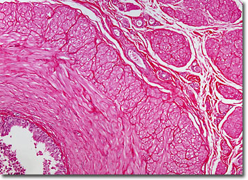
The ductus deferens, also known as the vas deferens, is a paired duct, or tube, that is part of the male reproductive system in mammals. Each ductus deferens extends from the epididymis to the pelvic region.
View an image of the ductus deferens section at 20x and 40x magnification.
Thick-walled, the ductus deferens is composed of several layers of tissue. Then innermost layer is a folded mucous membrane that is always moist. Encircling this layer are three layers of muscle tissue, which provide the tube with the ability to contract, a necessary action for the conveyance of bodily fluids and sperm. As the ductus deferens rises upwards from the epididymis towards the bladder, it also comes to be surrounded by nerve fibers, arteries, and veins, and the complex structure further encased in connective tissue is referred to as the spermatic cord. When the tube reaches the level of the bladder, however, it becomes separated from the connective tissue and extends across the top of the bladder and turns downward near the back of the structure, ending in an enlarged ampulla, which both stores and adds secretions to semen.
Severing the ductus deferens is the primary means of male sterilization for humans. The process, known as a vasectomy, is a relatively simple and safe surgical procedure that is often completed in less than 15 minutes. The ductus deferens is also sometimes the cause of natural sterility in men. Indeed, congenital bilateral absence of the vas deferens (CBAVD) is reportedly the cause of as many as 5 percent of all cases of male infertility. Associated with a mutated gene related to cystic fibrosis, the recessive condition may be passed onto offspring if a male with CBAVD utilizes medical procedures to enable him to have his own biological children.
BACK TO THE BRIGHTFIELD MICROSCOPY IMAGE GALLERY