
Brightfield Microscopy Digital Image Gallery
Amphibian Skin
Amphibians are cold-blooded animals that are generally considered the most primitive terrestrial members of the phylum Chordata. Frogs, toads, salamanders, newts, and caecilians are all members of this zoological class, each exhibiting a larval stage with gills that develops in an aquatic environment, external fertilization, and lungs in adulthood.
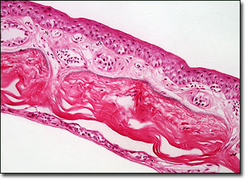
Due to their habitation of both terrestrial and freshwater locales at various stages in their life cycle, amphibians have a unique kind of permeable skin. Both oxygen and water are allowed to pass in and out of the delicate integument in order that homeostasis and a sufficient level of respiration may be maintained at all times, even when the animal is underwater. The skin of amphibians is also unusual because it must be kept in a relatively moist to help draw oxygen through the skin. In order to sustain the necessary moisture level, amphibians secrete mucus via glands contained in the skin. This mucous chiefly serves as a protective layer around the body when the animal is on land, but also facilitates a proper salt and water balance within the internal organs when the amphibian is submerged in water.
The skin of amphibians may exhibit a wide variety of colors and patterns. In many species, these characteristics of the skin help them blend into their natural environments in order to avoid predation. However, certain amphibians display bright warning colorations and patterns that may include hues of red, yellow, orange, and black, which serve as a bright signal to predators that they may be poisonous. Indeed, several species, such as the cane toads and poison arrow frogs, are equipped with skin glands that secrete powerful toxins.
BACK TO THE BRIGHTFIELD MICROSCOPY IMAGE GALLERY