
Frits Zernike
(1888-1966)
Frits Zernike was a Dutch mathematician and physicist who discovered the phase contrast phenomenon and was awarded the Nobel Prize in 1953. Zernike's parents were both teachers who specialized in mathematics, and his siblings also acquired good educations and prestige, with one brother becoming a physicist, a sister who married the well-known painter Jan Mankes, and another sister that rose to become one of Holland's foremost literary writers.
As a young man, Zernike was very interested in physics and chemistry. He accumulated a variety of spare equipment with which he would perform numerous experiments. Zernike was also interested in mathematics, astronomy, and photography, and conducted a number of investigations in these areas. He even dabbled in color photography when the field was largely experimental.
Zernike attended the University of Amsterdam in the early 1900s, where he majored in chemistry, with minors in mathematics and physics. While he was an undergraduate, he earned several gold medals for essays on probability and opalescence. Zernike eventually entered the graduate program and received a doctorate in physics in 1915.
He became an assistant to professor Kapteyn at Groningen University in 1913, and captured his first teaching assignment in mathematical physics at Groningen in 1915. As a researcher, Zernike flourished and studied a wide range of topics including correlation coefficients for molecules in a liquid, order-disorder theory, and optics.
It was Zernike's studies in optics that ultimately led to his Nobel Prize. He first received evidence of the phase contrast phenomenon in a study of diffraction gratings, when he was able to selectively detect transparent materials with different refractive indices. In 1938, Zernike built a microscope based on phase contrast illumination, but it initially received little attention. At the time, a lack of specimen contrast experienced with common microscopic techniques was one of the major concerns in optical microscopy.
Ironically, it was the German war machine that confiscated his invention and made a series of microscopes, which ultimately demonstrated the true utility of Zernike's technique. After the war, most microscope manufacturers rushed to produce microscopes with this enhanced mode of specimen illumination. Zernike was recognized by the Royal Microscopical Society and was also awarded the prestigious Rumford Metal by the Royal Society of London. His efforts were further rewarded by the Nobel committee with the prize in physics in 1953.
"How I Discovered Phase Contrast." Modified excerpts from Dr. Zernike's Nobel Prize address delivered in 1953 in Stockholm, Sweden and published in the March 11, 1955 issue of Science."I did not discover phase contrast while working with microscopes. Rather, it was my involvement with diffraction gratings that led to the breakthrough. Around 1930, our laboratory acquired a sizeable concave grating and the object's surface appeared striped due to periodic imperfections in the grating lines caused by the ruling machines of the time. Yet, when I focused a telescope on the surface from about six meters away, the stripes disappeared! A succession of experiments and calculations enabled me to explain the phenomenon. In a simpler instance, I found that a telescope with a vertical 2-millimeter slit placed close behind its objective could observe the diffraction pattern of a vertical line-source of light. The diffraction maxima could be observed, but their phases could not be discerned. The phases could be distinguished, however, if the diffraction image was projected onto a coherent background that could serve as a reference. I was familiar with Lord Rayleigh's simple process of making optically sound glass plate etchings using acid and utilized the technique to make what I call phase strips, glass plates with a single groove, one millimeter wide and etched to a depth of half a wavelength. When I placed a phase strip in the spectrum of the faulty grating and inspected it with a telescope, the stripes on the grating surface were clearly visible."
"As a physicist with an interest in optics it was not difficult for me to move from this subject to the microscope. Recall that Ernst Abbe's theoretical description of the microscope image associates the transparent object under a microscope with a grating. Abbe studied gratings consisting of alternating opaque and transparent strips (amplitude gratings), but I was more concerned with alternating thick and thin strips (phase gratings). Phase gratings generate spots of diffraction that demonstrate a phase difference of 90 degrees. For a phase object, when my phase strip is placed in the focal plane of the microscope objective, the direct image of the light source is brought into phase with the diffracted images of a phase object. The result is that the image viewed appears similar to that produced by an amplitude object. The image in the eyepiece of the microscope appears in black and white contrast, as if it were an absorbing object."
"I am impressed by the limitations of the human mind when I look back on these events. We are quick to learn-that is, to emulate what others have already done or thought-but slow to understand-that is, to realize the deeper connections. We are slowest of all, however, in conceiving new connections and in applying old ideas in a new area. In my situation, the truly new point was the fact that the diffraction pattern of the lines of gratings differ in phase from the principal line, and that the phases require projection of the diffraction image on a coherent background to be visualized. The full name of the microscopy technique could be something like phase-strip method for observing phase objects in good contrast, but I shortened it to phase contrast."
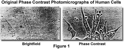
Presented in Figure 1 are photomicrographs of living tissue culture cells observed in brightfield illumination and phase contrast to demonstrate the enhanced contrast afforded by the latter technique. These images were taken over fifty years ago, and represent some of the original work done on phase specimens by Zernike and his colleagues
Introduction to Phase Contrast Microscopy - Phase contrast microscopy, first described in 1934 by Dutch physicist Frits Zernike, is a contrast-enhancing optical technique that can be utilized to produce high-contrast images of transparent specimens such as living cells, microorganisms, thin tissue slices, lithographic patterns, and sub-cellular particles (such as nuclei and other organelles). In effect, the phase contrast technique employs an optical mechanism to translate minute variations in phase into corresponding changes in amplitude, which can be visualized as differences in image contrast. One of the major advantages of phase contrast microscopy is that living cells can be examined in their natural state without being killed, fixed, and stained. As a result, the dynamics of ongoing biological processes in live cells can be observed and recorded in high contrast with sharp clarity of minute specimen detail.