
Brightfield Digital Image Gallery
Primate Hyaline Cartilage
As one of the three types of cartilage contributing to the primate skeletal structure, hyaline cartilage plays a role in endochondral ossification of bones. In primary ossification, masses of hyaline cartilage form models of future bones in the embryonic primate.
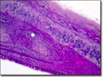
More common than elastic or fibrocartilage, hyaline cartilage contains collagen fibers arranged in a loose network. Occurring at free-moving bone joints and the ends of the ribs, this smooth cartilage is composed of chondrocytes, fiber, and mucopolysaccharides. It is also known as articular cartilage when arranged in thin, transparent sections covering the articular surfaces of bones and synovial joints. Articular cartilage is deformable, but quickly recovers its shape, making it well suited for reducing friction and absorbing shock in joints. Breakdown of this cartilage tissue due to repetitive loading or joint trauma leads to bone wear and pain associated with arthritis. The primate nose, trachea, bronchi, and larynx are composed of hyaline cartilage as well as other types of connective tissue and smooth muscle. Amphibians, reptiles and birds also utilize hyaline cartilage in their body structures.
The type of protein fiber in the cartilage matrix helps distinguish its type. In hyaline, the protein fibers are mostly collagen, which provides flexibility and support at the joints. Hyaline contains very little elastin compared to elastic cartilage. Unlike bone, cartilage is avascular. Since it is not served by the circulatory system, the chondrocytes of the cartilage must obtain nutrition by diffusion through the collagen gel matrix.
Contributing Authors
Cynthia D. Kelly, Thomas J. Fellers and Michael W. Davidson - National High Magnetic Field Laboratory, 1800 East Paul Dirac Dr., The Florida State University, Tallahassee, Florida, 32310.
BACK TO THE BRIGHTFIELD IMAGE GALLERY
BACK TO THE DIGITAL IMAGE GALLERIES
Questions or comments? Send us an email.
© 1995-2022 by Michael W. Davidson and The Florida State University. All Rights Reserved. No images, graphics, software, scripts, or applets may be reproduced or used in any manner without permission from the copyright holders. Use of this website means you agree to all of the Legal Terms and Conditions set forth by the owners.
This website is maintained by our
Graphics & Web Programming Team
in collaboration with Optical Microscopy at the
National High Magnetic Field Laboratory.
Last Modification Friday, Nov 13, 2015 at 02:19 PM
Access Count Since September 17, 2002: 16920
Visit the website of our partner in introductory microscopy education:
|
|