
Brightfield Digital Image Gallery
Fetal Elastic Cartilage
As with bone, elastic cartilage contains a matrix, fibers, and cells. In human elastic cartilage, the cartilage cells (chondrocytes) are contained in lacunae and the matrix contains abundant elastic fibers, explaining the characteristic flexibility of this type of connective tissue.
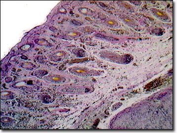
Providing flexible support to the human ears, the auditory canals, and the epiglottis, elastic cartilage is structurally identical to the more common hyaline cartilage except that it contains, in addition to Type II collagen fibers, a dense network of branching and anastomosing elastic fibers. Type II collagen fibers are composed predominantly of chondroitin sulfates and hyaluronic acid, with lesser amounts of keratin sulfate and herparin sulfate. Glycoproteins, the glue that holds the matrix together, include link proteins, fibronectin, chondronectin, and tissue fluid. At the core of the elastic cartilage mass, the network is at its densest. Surrounding and nourishing the elastic cartilage mass is the perichondrium, and unlike hyaline cartilage, the chondrocytes are found in isogenous groups.
Elastic cartilage develops from a primitive type of connective tissue, which contains wavy bundles of fibrils that are neither elastin nor collagen. Fibroblasts secrete elastin in the fetus and the fiber bundles branch, and are cross-linked, creating a gel-like consistency. As with hyaline cartilage, the fetal tissue chondrocytes differentiate with the chondroblasts at the center, first secreting matrix material, which then separates into the individual cells. During embryonic development, mesenchymal cells retract their cytoplasmic extensions and assume the rounded shape of chondroblasts. As they become tightly packed, the increased cell-to-cell contact stimulates cartilage differentiation, progressing from the center outwards to the periphery of the fetal tissue.
Contributing Authors
Cynthia D. Kelly, Thomas J. Fellers and Michael W. Davidson - National High Magnetic Field Laboratory, 1800 East Paul Dirac Dr., The Florida State University, Tallahassee, Florida, 32310.
BACK TO THE BRIGHTFIELD IMAGE GALLERY
BACK TO THE DIGITAL IMAGE GALLERIES
Questions or comments? Send us an email.
© 1995-2022 by Michael W. Davidson and The Florida State University. All Rights Reserved. No images, graphics, software, scripts, or applets may be reproduced or used in any manner without permission from the copyright holders. Use of this website means you agree to all of the Legal Terms and Conditions set forth by the owners.
This website is maintained by our
Graphics & Web Programming Team
in collaboration with Optical Microscopy at the
National High Magnetic Field Laboratory.
Last Modification Friday, Nov 13, 2015 at 02:19 PM
Access Count Since September 17, 2002: 17128
Visit the website of our partner in introductory microscopy education:
|
|