
Brightfield Digital Image Gallery
Dicot Leaf Epidermis
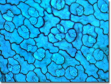
The dicot plant genus Sedum includes several hundred species, which are generally classified as succulents, and which have thick leaves able to withstand a drought. The digital image below illustrates a stained thin section of Sedum dicotyledon epidermal cells with numerous stomata, each having guard cells encircled by subsidiary cells.
Leaves, which are present on almost all vascular plants, are a main appendage of the stem and represent the principal site of photosynthesis. The leaves found on common plants are highly varied, and differ to a significant degree in shape and structure depending primarily on habitat. Leaf shape, margins, tip geometry, and vein patterns are characteristics that are utilized as an aid in identifying the different species of flowering plants. Most leaves have several common features, including a blade (often termed a lamina), which is the flattened part of the leaf structure. Leaves are typically equipped with a petiole, or stalk, employed to connect the leaf to the plant stem, and those that are absent this feature are termed sessile. Another common trait found in some plants is the presence of stipules, which are small leaf-shaped growths near the base of the stalk.
The vein patterns exhibited by leaves are important in distinguishing between monocots and dicots. In general, dicot leaves have either a pinnate or palmate vein pattern with netted veins and a large mid-vein, whereas monocot leaves usually have parallel veins. The blades of leaves may be simple, dissected, or compound.
Contributing Authors
Cynthia D. Kelly, Thomas J. Fellers and Michael W. Davidson - National High Magnetic Field Laboratory, 1800 East Paul Dirac Dr., The Florida State University, Tallahassee, Florida, 32310.
BACK TO THE BRIGHTFIELD IMAGE GALLERY
BACK TO THE DIGITAL IMAGE GALLERIES
Questions or comments? Send us an email.
© 1995-2025 by Michael W. Davidson and The Florida State University. All Rights Reserved. No images, graphics, software, scripts, or applets may be reproduced or used in any manner without permission from the copyright holders. Use of this website means you agree to all of the Legal Terms and Conditions set forth by the owners.
This website is maintained by our
Graphics & Web Programming Team
in collaboration with Optical Microscopy at the
National High Magnetic Field Laboratory.
Last Modification Friday, Nov 13, 2015 at 01:19 PM
Access Count Since September 17, 2002: 30776
Visit the website of our partner in introductory microscopy education:
|
|