
Human Vision and Color Perception
Human vision is a complex process that is not yet completely understood, despite hundreds of years of study and research. The complex physical process of visualizing something involves the nearly simultaneous interaction of the eyes and the brain through a network of neurons, receptors, and other specialized cells.
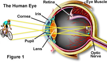
The human eye is equipped with a variety of optical elements including the cornea, iris, pupil, a variable-focus lens, and the retina, as illustrated above in Figure 1. Together, these elements work to form images of the objects in a person's field of view. When an object is observed, it is first focused through the cornea and lens onto the retina, a multilayered membrane that contains millions of light-sensitive cells that detect the image and translate it into a series of electrical signals. These image capturing receptors of the retina are termed rods and cones, and are connected with the fibers of the optic nerve bundle through a series of specialized cells that coordinate the transmission of the electrical signals to the brain. In the brain, the optic nerves from both eyes join at the optic chiasma where information from their retinas is correlated. The visual information is then processed through several steps, eventually arriving at the visual cortex, which is located on the lower rear section of each half of the cerebrum.
| Interactive Tutorial | |||||||||||
|
|||||||||||
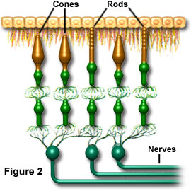
A particularly specialized component of the eye is the fovea centralis, which is located on the optical axis of the eye in an area near the center of the retina. This area exclusively contains high-density tightly packed cone cells and is the area of sharpest vision. The density level of cone cells decreases outside of the fovea centralis and the ratio of rod cells to cone cells gradually increases. At the periphery of the retina, the total number of both types of light receptors decreases substantially, causing a dramatic loss of visual sensitivity at the retinal borders. This is offset, however, by the fact that humans constantly scan objects in their field of view, usually resulting in a perceived image that is uniformly sharp. A graphical illustration of the spatial arrangement of rod and cone cells and their connection to neurons within the retina can be seen below in Figure 2.
Rod cells are most sensitive to green wavelengths of light (about 550-555 nanometers), although they display a broad range of response throughout the visible spectrum. They are the most populous visual receptor cells in humans, each eye containing about 130 million rods. Extremely responsive, the light sensitivity of rod cells is about 1000 times that of cone cells. However, the images generated by rod stimulation alone are relatively unsharp and confined to shades of gray, similar to those found in a black and white soft-focus photographic image. Rod vision is commonly referred to as scotopic or twilight vision because in low light levels it enables individuals to distinguish shapes and the relative brightness of objects, but not their colors.
Cones, on the other hand, consist of three different types of cells, each "tuned" to a distinct wavelength peak of response centered at either 430, 535, or 590 nanometers. Often referred to as photopic vision, cone vision is dominant at normal light levels, both indoors and out. The quantity of cone cells possessed by humans, however, is much smaller than the number of rod cells, each eye only containing about 7 million cones. Stimulation of these visual receptors results in what is known as true color vision.
The relative intensity of the stimulation incurred by each of the three types of cone receptors is what largely determines which color is imaged. For example, a beam of light that contains mostly blue short-wavelength radiation stimulates the cone cells that respond to 430-nanometer light far more than the other two cone types and, therefore, that light is seen as blue. Correspondingly, light with a majority of wavelengths centered around 550 nanometers appears green, and a beam containing mostly 600 nanometer wavelengths or longer is seen as red. When all three types of cone cells are stimulated equally, light is perceived as being achromatic or white. For instance, noon sunlight appears to humans as white light because it contains approximately equal amounts of red, green, and blue light, uniformly stimulating all types of cone receptors.
Normal cones and pigment sensitivity enable humans to distinguish all of the different colors as well as subtle mixtures of hues. This type of color vision is known as trichromacy and relies upon the mutual interaction of all three types of photoreceptor cones. Yet, human color perception is also dependent upon illumination levels, shifts in color sensitivity occurring whenever lighting is varied. For example, blue colors look relatively brighter in dim light, while red colors appear more vivid in bright light. This effect can be simply observed by pointing a flashlight onto a color print, which results in the reds suddenly appearing much brighter and more saturated.
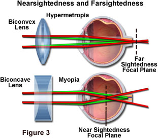
Focus in the eye is controlled by a combination of elements, including the iris, lens, cornea, and muscle tissue. Properly functioning together, these components can alter the shape of the lens so that the eye can focus on both nearby and distant objects. However, in some instances the components do not work correctly or the eye is slightly altered in shape and the focal point does not intersect with the retina. As people age, for instance, the lenses of their eyes become harder and cannot be focused properly, which results in poor vision. If the point of an eye's focus is short of the retina, as illustrated in the lower part of Figure 3, the condition is called nearsightedness or myopia. People with this affliction are unable to focus on distant objects. In cases where the eye's focal point is behind the retina, as illustrated in the upper part of Figure 3, people have trouble focusing on nearby objects, which is a condition called hypermetropia, commonly known as farsightedness. These malfunctions of the eye can usually be corrected through the use of glasses, with concave lenses correcting myopia and convex lenses rectifying hypermetropia.
Contributing Authors
Mortimer Abramowitz - Olympus America, Inc., Two Corporate Center Drive., Melville, New York, 11747.
Shannon H. Neaves and Michael W. Davidson - National High Magnetic Field Laboratory, 1800 East Paul Dirac Dr., The Florida State University, Tallahassee, Florida, 32310.