
Enhanced Brightfield Microscopy
The default illumination mode for the Intel Play QX3 Computer Microscope is either transmitted or reflected brightfield. When the lamp housed in the microscope body is turned on, light from the bulb passes through a frosted diffusion screen and is projected onto the surface of an opaque specimen such as a coin, insect, paper clip, etc.
The light striking the opaque specimen is then reflected, refracted, and diffracted into the front lens of the objective where an image of the specimen is formed. This is termed Reflected Light Illumination microscopy because light entering the objective is first reflected from the surface of the opaque specimen. The light is called Brightfield because it consists essentially of unfiltered 3200K tungsten-balanced illumination. There are no polarizers, retardation plates, phase plates, Wollaston prisms, or colored Rheinberg gels that alter the properties of white light.
Alternatively, when the stage lamp is turned on, the body lamp is automatically and simultaneously turned off, and light passes through the stage diffusion filter, around and through the specimen and into the objective front lens (Figure 1). This light is termed Transmitted Light Illumination because light entering the objective is first transmitted through a transparent specimen. It is also referred to as brightfield illumination for the same reasons discussed for reflected light.
Brightfield illumination from either of the QX3 microscope bulbs is of sufficient intensity to illuminate a majority of the simple specimens that children are likely to image with the microscope at low (10x) magnifications. As the objective magnification is ratcheted up to 60x and 200x, light intensity progressively decreases to a point where many specimens become poorly illuminated and the digital images obtained from the software become very dark. To remedy this situation, we have devised several methods to improve brightfield illumination using both transmitted and reflected light modes.
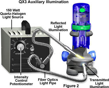
The best form of auxiliary illumination, both for transmitted and reflected light microscopy, is that provided by quartz-halogen illuminators designed for use with flexible fiber optic light guides or pipes (illustrated in Figure 2). These light sources provide long lasting high intensity illumination, but with the undesired side effect of also producing a significant amount of infrared light in the form of heat. The illuminators generally provide an air or convection cooling mechanism for the internal bulb to increase its life span and to keep the housing as cool as possible. Unfortunately, direct light from this high intensity source can result in heat absorption by an object positioned on the microscope stage.
Light from the quartz-halogen bulb is directed onto the microscope stage by the flexible fiber optic light pipes or guides that can be positioned in an almost infinite number of different configurations. These guides are quite helpful in solving the heat problem, because their internal optical fibers transmit primarily visible illumination with minimal transmission in the near infrared or near ultraviolet spectrum. The result is an intense illumination centered in the visible light spectrum without the associated infrared heat output that can alter or damage either the specimen or the microscope itself.
Light intensity is controlled by a potentiometer housed on the faceplate of the illuminator. The voltage delivered to the lamp can be varied with the potentiometer in ranges from zero current to 120 volts in continuous increments. Light emitted from the quartz-halogen light source has a constant color temperature that remains at 3200K throughout the adjustable voltage range, producing a very stable illumination source.
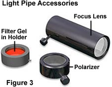
The light source illustrated in Figure 2 is a Dolan-Jenner model number 180 illuminator that costs about $500 when equipped with the dual light pipes shown in the figure. Optical fibers in the light pipes are terminated at the end of the pipes and produce a large, diffuse cone of light. This cone of light can be concentrated using lenses (Figure 3) that fit over the end of the light pipe and focus the light to various degrees depending upon the focal length of the lens. Accessories for light pipe lenses include polarizers, which will greatly reduce light intensity, and holders for colored filter gels that will add specific colors to the light (Figure 3).
|
||||||||||||||||||||||||||||||||||||||||||||||||||||||
We have measured the illumination intensity of the QX3 microscope body and stage illuminators for the purposes of comparison to the auxiliary external quartz-halogen illuminator. The QX3 body illuminator produces light measuring approximately 0.0165 lumens, while the substage lamp emits less light that we have measured to be approximately 0.0046 lumens. The difference between these two light sources arises from the mixing chamber built within the microscope stage. Light entering the mixing chamber is bounced around and up through a frosted diffusion screen that is 3 centimeters in diameter and greatly reduces the light density.
The light intensity of the quartz-halogen fiber optic light pipes has also been measured and the results are presented in Table 1. At full intensity, the quartz-halogen illuminator produces approximately four times the illumination of the QX3 body lamp and about 16 times the illumination of the stage lamp. When a focusing lens is added, these numbers are doubled. Conversely, when a polarizer is attached to a light pipe containing a focusing lens, illumination intensity is reduced to about half that of the light pipe alone. These numbers make it clear that addition of the external auxiliary illumination greatly enhances the microscope's ability to produce digital images, especially at the higher magnifications.
When highly reflective surfaces are imaged with auxiliary oblique illumination, glare can become a problem in digital images. To help alleviate this problem, polarizers can be attached to the light pipe focusing lens(es) to reduce the amount of polarized light reflected due to the Brewster effect. Orient the polarizers with their vibration planes positioned parallel to the optical axis of the microscope so that light passed through to the specimen surface has a polarization vector perpendicular to the polarized light that is reflected at the Brewster angle. This will decrease the amount of glare in digital images recorded with the QX3 software.
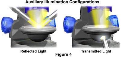
Close-up illustrations for auxiliary illumination using quartz-halogen illuminator light pipes for both transmitted and reflected light microscopy are presented in Figure 1. The configuration on the left uses twin light pipes from both sides of the QX3 microscope for maximum illumination potential. This method of illumination was used for all of the reflected light digital photomicrographs in our QX3 Digital Image Gallery.
To configure the microscope for transmitted illumination, we drill a quarter-inch hole in the base of the mixing chamber and point a light pipe directly into the hole to project light on the rear wall (painted white) of the mixing chamber. This approach has produced the most even illumination for transmitted light. Using this technique, we have successfully been able to obtain acceptable digital images of stained thin sections of plant and animal tissues.
To add color to digital images made with the QX3 microscope, use colored gels with the filter holder attachment for the light pipes illustrated in Figure 3. Depending upon the optical density of the dyes used to fabricate the gels, the light intensity will fall somewhat when gels are added. We have measured the illumination intensity using the four most common colors: red, green, blue, and yellow. The results are presented in Table 2.
|
|||||||||||||||||||||||||||||||||||||||||||||||||||||||||||||||||||
It is evident from the data in Table 2 that the green filter has the greatest optical density and passes the least amount of light, while the yellow filter has the least density and passes the greatest amount of light. Colored gels are very useful in adding color using the reflected light Rheinberg illumination technique with both transparent and opaque specimens. If two light pipes are being used with different colored gels, it is important to ensure that the optical densities of both filters are approximately matched, otherwise the filter with least density will far overpower the one with the most optical density and its color will saturate the specimen.
Quartz-halogen light sources are expensive and unavailable to most children. There are a number of good alternatives to auxiliary illumination, which are available at most department stores. High-intensity desk lamps are usually very compact and can be useful in providing auxiliary illumination.
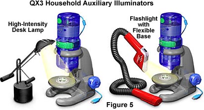
The QX3 microscopes illustrated in Figure 5 are receiving assistance by common household auxiliary illumination sources. The high-intensity Tensor-style desk lamp is equipped with a low wattage tungsten-halide lamp that has a color spectrum very similar to the quartz-halogen illuminator and the native QX3 lamps. Many varieties of these lamps have adjustable voltages that allow the user to select between a limited number of intensity choices. Desk lamps are perfectly adequate for a number of applications that take advantage of enhanced reflected brightfield illumination at a fraction of the cost of the quartz-halogen systems. Because of the diffuse nature of the illumination provided by these lamps, they are of little use for transmitted light microscopy.
The QX3 microscope can be adapted for transmitted light using the common Mini Maglite flashlights available in most department stores. These miniature flashlights have a 2 centimeter reflector and will produce concentrated illumination when pointed into a quarter- to half-inch hole drilled in the substage mixing chamber. There are probably other flashlight models that can be substituted for the Mini Maglite when using this technique.
Finally, the common Snakelite flashlights, with a flexible base, provide a convenient and easy method for producing auxiliary illumination for reflected light microscopy. Although these flashlights do not provide the illumination intensity of their more expensive counterparts, they are widely available and usually cost less than $20 in most department stores.
It is important to remember that providing proper specimen illumination is the key to high-quality digital imaging with an optical microscope. Auxiliary illumination, regardless of the source, will improve the performance of the Intel Play QX3 microscope and allow anyone to produce the best quality digital images that are possible with the limited optics of this microscope.
BACK TO INTEL QX3 SPECIALIZED TECHNIQUES
Questions or comments? Send us an email.
© 1995-2025 by Michael W. Davidson and The Florida State University. All Rights Reserved. No images, graphics, software, scripts, or applets may be reproduced or used in any manner without permission from the copyright holders. Use of this website means you agree to all of the Legal Terms and Conditions set forth by the owners.
This website is maintained by our
Graphics & Web Programming Team
in collaboration with Optical Microscopy at the
National High Magnetic Field Laboratory.
The QX3 microscope design is copyrighted © 2025 by Mattel, Inc. Intel® Play™ is a registered trademark of the Intel Corporation. These companies reserve all of their rights and privileges under copyright law. The material contained in this website is solely the opinion of the authors and is intended for eduational use only.
Last Modification Friday, Nov 13, 2015 at 02:18 PM
Access Count Since December 24, 1999: 44360
Visit the official Intel Play website:
![]()