
Euplotes (Protozoa) Movies
Euplotes Video No. 1 - A ventral (belly) view of a euplotes shows its internal structures and a waving band of cirri, just before it moves out of field; at a magnification of 200x with a playing time of 40.9 seconds. Choose a playback format that matches your connection speed: 28.8k (modem), 56.6k (modem), or T1/Cable/DSL, or download this video clip in MPEG format (4.39 MB).
Euplotes Video No. 2 - A close-up view of a euplotes shows the band of cirri that it uses for locomotion; under phase contrast illumination at a magnification of 400x with a playing time of 19.5 seconds. Choose a playback format that matches your connection speed: 28.8k (modem), 56.6k (modem), or T1/Cable/DSL, or download this video clip in MPEG format (5.67 MB).
Euplotes Video No. 3 - An individual euplotes, its cirri and tactile cilia waving, along with other microorganisms; under phase contrast illumination at a magnification of 200x with a playing time of 20.3 seconds. Choose a playback format that matches your connection speed: 28.8k (modem), 56.6k (modem), or T1/Cable/DSL, or download this video clip in MPEG format (4.78 MB).
Euplotes Video No. 4 - A pair of joined Euplotes organisms, either completing asexual reproduction or undergoing conjugation - exchanging nuclear material; under phase contrast illumination at a magnification of 100x with a playing time of 20.0 seconds. Choose a playback format that matches your connection speed: 28.8k (modem), 56.6k (modem), or T1/Cable/DSL, or download this video clip in MPEG format (8.69 MB).
Euplotes Video No. 5 - An individual euplotes, its cirri and tactile cilia waving, along with other microorganisms; under phase contrast illumination at a magnification of 200x with a playing time of 17.4 seconds. Choose a playback format that matches your connection speed: 28.8k (modem), 56.6k (modem), or T1/Cable/DSL, or download this video clip in MPEG format (3.82 MB).
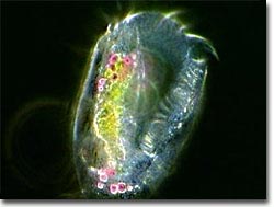
Euplotes Video No. 6 - A close-up view of a euplotes shows its internal structures and a waving band of cirri; under darkfield illumination at a magnification of 400x with a playing time of 39.4 seconds. Choose a playback format that matches your connection speed: 28.8k (modem), 56.6k (modem), or T1/Cable/DSL, or download this video clip in MPEG format (8.79 MB).
Euplotes Video No. 7 - A pair of joined Euplotes organisms, either completing asexual reproduction or undergoing conjugation - exchanging nuclear material; under phase contrast illumination at a magnification of 100x with a playing time of 14.4 seconds. Choose a playback format that matches your connection speed: 28.8k (modem), 56.6k (modem), or T1/Cable/DSL, or download this video clip in MPEG format (4.36 MB).
Euplotes Video No. 8 - An encounter with another microorganism sends this euplotes scampering; under phase contrast illumination at a magnification of 200x with a playing time of 4.6 seconds. Choose a playback format that matches your connection speed: 28.8k (modem), 56.6k (modem), or T1/Cable/DSL, or download this video clip in MPEG format (1.12 MB).