
Wim van Egmond
Odontella Diatoms
Diatoms are unicellular organisms that secrete overlapping siliceous cell walls called frustules. The often-beautiful organisms have an extensive fossil record that dates back to the Cretaceous period, some 144 to 66.4 million years ago.
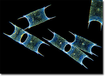
Most diatoms are free-living, planktonic species, but some live in extensive colonies or linked into tiny chains, like many members of the genus Odontella. These organisms frequently feature long spines on their frustules and may stay connected to one another after cell division via a crossing of the spines. Other items that may be exhibited on diatom frustules include intricate patterns composed of ridges, round perforations, linear markings, ribs, and vertical canals. The beauty of diatom designs has earned them the well-deserved title “jewels of the sea.”
Diatoms are not only pleasing to the eye, but are extremely useful. Estimates indicate that approximately 25 percent of all organic carbon fixation on the planet is initiated by diatoms, and the microscopic organisms are also an important food source to a great number of aquatic creatures. Interestingly, diatoms are so functional that they are even utilized by humans after they are dead. A mineral substance called diatomite or diatomaceous earth is composed of the skeletons of ancient diatoms that settled on the bottom of the oceans or along lakebeds. Humans mine the chalky material from large deposits in states such as California and Nevada and use it for filtration purposes, as an insulator, and as a component in an array of products, including metal polish, detergents, and even toothpaste.
BACK TO WIM VAN EGMOND GALLERY