
Wim van Egmond
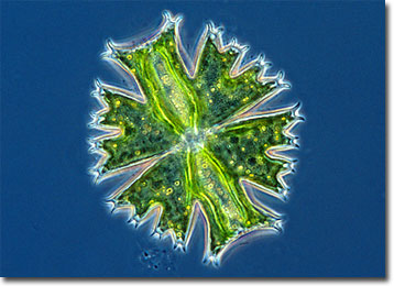
Micrasterias crux-melitensis
Desmids are microscopic green algae that may occur as unicells or in filaments. The tiny organisms are particularly interesting subjects to microscopists because they exhibit remarkable variation in their ornamentation and cell shape.
The term “desmid” is derived from the Greek “desmos,” meaning “bond” or “chain.” The name originated at a time when the organisms were believed to consist of pairs of joined cells. However, desmids have since been discovered to usually be composed of a single cell that is constricted in the center so that two symmetrical semicells connected by an isthmus are created. The nucleus of a desmid is typically contained in the isthmus, and inside each semicell are one or more starch-storing pyrenoids and chloroplasts, which facilitate photosynthesis. Reproduction of desmids may involve the sexual act of conjugation or may take place asexually, individuals dividing along the isthmus and each half developing a new semicell.
The name of the genus Micrasterias means “small star,” a fitting moniker due to the typical shape of its members. The organisms, which are considered placoderm desmids since they feature cell walls composed of two sections that attach in the mid-region, are flat and variously lobed in beautiful stellar-like designs. The more than 40 known species of Micrasterias may be further ornamented with patterns of pores, spines, granules, or other protuberances. Most often found in acidic waters and bogs, the organisms may grow between 80 to 200 micrometers in diameter. A typical species is Micrasterias crux-melitensis, which primarily inhabits moorlands and is approximately 102 to 129 micrometers long.
BACK TO WIM VAN EGMOND GALLERY