
Plant Cell Wall
One of the most important distinguishing features of plant cells is the presence of a cell wall. The relative rigidity of the cell wall renders plants sedentary, unlike animals, whose lack of this type of structure allows their cells more flexibility, which is necessary for locomotion. The plant cell wall serves a variety of functions. Along with protecting the intracellular contents, the structure bestows rigidity to the plant, provides a porous medium for the circulation and distribution of water, minerals, and other nutrients, and houses specialized molecules that regulate growth and protect the plant from disease.
Cell walls are significantly thicker than plasma membranes and were visible even to early microscopists, including Robert Hooke, who originally identified the structures in a sample of cork, and then coined the term cells in the 1660s. The thickness, as well as the composition and organization, of cell walls can vary significantly. Many plant cells have both a primary cell wall, which accommodates the cell as it grows, and a secondary cell wall they develop inside the primary wall after the cell has stopped growing. The primary cell wall is thinner and more pliant than the secondary cell wall, and is sometimes retained in an unchanged or slightly modified state without the addition of the secondary wall, even after the growth process has ended.
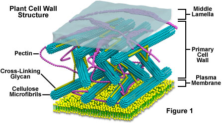
The main chemical components of the primary plant cell wall include cellulose (in the form of organized microfibrils; see Figure 1), a complex carbohydrate made up of several thousand glucose molecules linked end to end. In addition, the cell wall contains two groups of branched polysaccharides, the pectins and cross-linking glycans. Organized into a network with the cellulose microfibrils, the cross-linking glycans increase the tensile strength of the cellulose, whereas the coextensive network of pectins provides the cell wall with the ability to resist compression. In addition to these networks, a small amount of protein can be found in all plant primary cell walls. Some of this protein is thought to increase mechanical strength and part of it consists of enzymes, which initiate reactions that form, remodel, or breakdown the structural networks of the wall. Such changes in the cell wall directed by enzymes are particularly important for fruit to ripen and leaves to fall in autumn.
The secondary plant cell wall, which is often deposited inside the primary cell wall as a cell matures, sometimes has a composition nearly identical to that of the earlier-developed wall. More commonly, however, additional substances, especially lignin, are found in the secondary wall. Lignin is the general name for a group of polymers of aromatic alcohols that are hard and impart considerable strength to the structure of the secondary wall. Lignin is what provides the favorable characteristics of wood to the fiber cells of woody tissues and is also common in the secondary walls of xylem vessels, which are central in providing structural support to plants. Lignin also makes plant cell walls less vulnerable to attack by fungi or bacteria, as do cutin, suberin, and other waxy materials that are sometimes found in plant cell walls.
A specialized region associated with the cell walls of plants, and sometimes considered an additional component of them, is the middle lamella (see Figure 1). Rich in pectins, the middle lamella is shared by neighboring cells and cements them firmly together. Positioned in such a manner, cells are able to communicate with one another and share their contents through special conduits. Termed plasmodesmata, these small passages penetrate the middle lamella as well as the primary and secondary cell walls, providing pathways for transporting cytoplasmic molecules from one cell to another.