
Peroxisomes
Microbodies are a diverse group of organelles that are found in the cytoplasm of almost all cells, roughly spherical, and bound by a single membrane. There are several types of microbodies, including lysosomes, but peroxisomes are the most common. All eukaryotes are comprised of one or more cells that contain peroxisomes. The organelles were first discovered by the Belgian scientist Christian de Duve, who also discovered lysosomes.
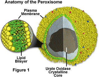
Peroxisomes contain a variety of enzymes, which primarily function together to rid the cell of toxic substances, and in particular, hydrogen peroxide (a common byproduct of cellular metabolism). These organelles contain enzymes that convert the hydrogen peroxide to water, rendering the potentially toxic substance safe for release back into the cell. Some types of peroxisomes, such as those in liver cells, detoxify alcohol and other harmful compounds by transferring hydrogen from the poisons to molecules of oxygen (a process termed oxidation). Others are more important for their ability to initiate the production of phospholipids, which are typically used in the formation of membranes.
In order to carry out their activities, peroxisomes use significant amounts of oxygen. This characteristic of the organelles would have been extremely important millions of years ago, before cells contained mitochondria, when the Earth's atmosphere first began to amass large amounts of oxygen due to the actions of photosynthetic bacteria. Peroxisomes would have been primarily responsible at that time for detoxifying cells by decreasing their levels of oxygen, which was then poisonous to most forms of life. The organelles would have provided the cellular benefit of carrying out a number of advantageous reactions as well. Later, when mitochondria eventually evolved, peroxisomes became less important (in some ways) to the cell since mitochondria also utilize oxygen to carry out many of the same reactions, but with the additional benefit of generating energy in the form of adenosine triphosphate (ATP) at the same time.
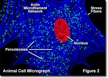
Peroxisomes are similar in appearance to lysosomes, another type of microbody, but the two have very different origins. Lysosomes are generally formed in the Golgi complex, whereas peroxisomes self-replicate. Unlike self-replicating mitochondria, however, peroxisomes do not have their own internal DNA molecules. Consequently, the organelles must import the proteins they need to make copies of themselves from the surrounding cytosol. The importation process of peroxisomes is not yet well understood, but it appears to be heavily dependent upon peroxisomal targeting signals composed of specific amino acid sequences. These signals are thought to interact with receptor proteins present in the cytosol and docking proteins present in the peroxisomal membrane. As more and more proteins are imported into lumen of a peroxisome or are inserted into its membrane, the organelle gets larger and eventually reaches a point where fission takes place, resulting in two daughter peroxisomes. Illustrated in Figure 2 is a fluorescence digital image of an African water mongoose skin fibroblast cell stained with fluorescent probes targeting the nucleus (red), actin cytoskeletal network (blue), and peroxisomes (green).
Since the early 1980s, a number of metabolic disorders have been discovered to be caused by molecular defects in peroxisomes. Two major categories have been described so far. The first category consists of disorders of peroxisome biogenesis in which the organelle fails to develop normally, causing defects in numerous peroxisomal proteins. The second category involves defects of single peroxisomal enzymes. Studies indicate that approximately one in every 20,000 people has some type of a peroxisomal disorder. The most serious of these disorders is Zellweger syndrome, which is characterized by an absence or reduced number of peroxisomes in the cells. Present in patients at birth (congenital), Zellweger syndrome has no cure or effective treatment and usually causes death within the first year of life.