
The Nucleolus
The nucleolus is a prominent sub-nuclear structure that is not bound by a membrane and resides within the nuclear matrix. Though known to exist since the eighteenth century, the primary function of the nucleolus was not discovered until the 1960s. It is now been determined that nucleoli manufacture the subunits that combine to form ribosomes, the cell's protein-producing factories. Accordingly, the size of nucleoli depends upon the ribosomal requirements of the type of cell in which they are found. In cells that produce large amounts of protein, and thus call for significant numbers of ribosomes, the size of the nucleolus is considerable, sometimes occupying as much as 25 percent of the total volume of the nucleus.
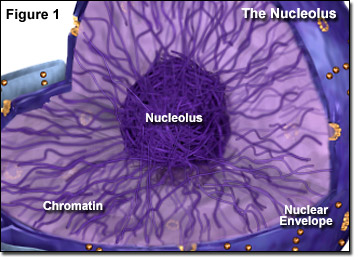
Through the microscope, the nucleolus appears like a large dark spot within the nucleus (see Figure 2). Eukaryotic cells often contain a single nucleolus, but several are also possible. The exact number of nucleoli is fixed among members of the same species. Each diploid cell in the human body features only one nucleolus, though immediately after cell division ten tiny nucleoli appear before they coalesce into a single, large nucleolus. This is because nucleoli are formed at certain chromosome sites usually referred to as nucleolus organizer regions (NORs), and two copies of five different human chromosomes contain NORs. The DNA found at chromosomal NORs encodes the genes for ribosomal RNA (rRNA). At the onset of mitosis, the single nucleolus present in a human cell disappears, and subsequent to the process, the formation of the new nucleolus, which is created from the ten smaller nucleolus-like structures that develop from the NORs, can be observed.
The nucleolus is comprised of granular and fibrillar components, as well as an ill-defined matrix, in addition to DNA. The granular material consists of ribosomal subunits that have already been formed but have not yet matured and are waiting to be exported to the cytoplasm. The threadlike fibrillar part of a nucleolus is predominantly composed of rRNA molecules and associated proteins that have joined together to form fibrils. It has not yet been determined exactly how the various components of the nucleolus are secured together and organized.
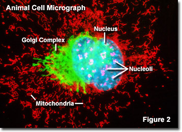
Illustrated in Figure 2 is a fluorescence digital image of a Swiss mouse embryo fibroblast cell stained with fluorescent probes targeting the nucleus (blue), mitochondria network (red), Golgi complex (green) and nucleoli (magenta) to demonstrate the proximity of these structures. The nucleoli of this cell were targeted with an antibody to fibrillarin, a small nuclear ribonucleoprotein (SnRNP) involved in ribosomal RNA processing and centralized within the sub-organelle. Fibrillarin has a well conserved amino acid sequence that enables the protein to serve as an excellent marker for nucleoli in a wide variety of species.
In recent years, scientists have been industriously investigating a possible connection between the nucleolus and cell senescence (aging). Most evidence for such a link thus far has come from studies with yeast cells, some of which suggest that cumulative damage to ribosomal RNA genes and fragmentation of the nucleolus may be central to the aging process. Support for the theory has also come from genetic research focused upon Werner syndrome, an inherited disease characterized by premature aging, but additional studies are needed to more precisely understand any possible role the nucleolus plays in senescence.