
Leaf Tissue Organization
The plant body is divided into several organs: roots, stems, and leaves. The leaves are the primary photosynthetic organs of plants, serving as key sites where energy from light is converted into chemical energy. Similar to the other organs of a plant, a leaf is comprised of three basic tissue systems, including the dermal, vascular, and ground tissue systems. These three motifs are continuous throughout an entire plant, but their properties vary significantly based upon the organ type in which they are located. All three tissue systems are illustrated in Figure 1, which is a cutaway drawing of a typical leaf.
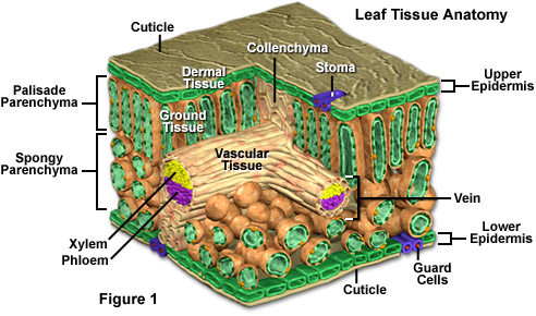
The dermal tissue of a plant, more specifically referred to as the epidermis, is an outer protective layer of typically polygonal cells, which helps defend against injury and invasion by foreign organisms. The epidermis of the leaf also functions in a more specialized manner by secreting a waxy substance that forms a coating, termed the cuticle, on the surface of the leaf. An adaptation unique to terrestrial plants, the cuticle functions chiefly in the retention of water. As presented in Figure 1, the cells that comprise the epidermis of a leaf are arranged very tightly together in a single stratum.
Microscopic pores known as stomata are the only breaches in the otherwise continuous layer of the leaf epidermis. Each individual pore, or stoma, is, in fact, a small opening between a pair of specialized cells known as guard cells. By modifying the size of the stomata, guard cells are able to regulate gas exchange and transpiration. Such modifications are influenced by various environmental factors. For example, when the weather is unusually hot and dry, the guard cells of plants in danger of losing too much water narrow the stomata width in order to reduce evaporation from the leaf interior.
In order for leaves to obtain water and minerals from the roots and for food manufactured in mature leaves to be transported to the roots and other nonphotosynthetic regions, each leaf must be connected to the overall vascular structure of the plant. Accordingly, the main vascular bundle of xylem and phloem present in the stem of a plant bifurcates into leaf traces, which are branches of vascular tissue that supply leaves. Each leaf trace further branches into the familiar veins that can often be seen along the surface of leaves, and the veins repeatedly subdivide as well. The vascular components, which serve as a basic skeletal structure in addition to functioning in the transport of materials, extend throughout the mesophyll so that the xylem and phloem are brought into propinquity with leaf tissues that carry out photosynthesis.
The mesophyll is the mid-section of a leaf, located between the upper and lower epidermal layers. Not only is vasculature found in the mesophyll, but also the ground tissue of a leaf. Ground tissue comprises the bulk of a plant leaf and is generally comprised of a variety of cell types, the predominant of which are parenchyma. Often less specialized than other plant cell types, parenchyma cells are surrounded by thin, flexible primary walls and execute most of the plant’s metabolic activities. The parenchyma cells present in leaves contain chloroplasts, which are the sites of photosynthesis.
In Figure 1, the mesophyll is divided into two conspicuously different regions, a characteristic common among the leaves of many dicotyledons. The upper section is termed the palisade parenchyma and consists chiefly of elongated columnar parenchyma cells that contain three to five times the number of chloroplasts as the cells that comprise the lower layer, known as the spongy parenchyma. The cells of the spongy parenchyma are irregularly shaped, allowing gases to circulate through the numerous air spaces between them to the palisade parenchyma. The stomata, which are particularly important for gas exchange, tend to be surrounded by exceptionally large air spaces.
A small group of collenchyma cells are also illustrated in the mesophyll of the leaf section presented in Figure 1. As depicted, collenchyma cells occur in aggregates just beneath the epidermis and possess thicker primary cell walls than parenchyma cells. The thickness of the walls, however, does exhibit notable variation. The main function of collenchyma cells is to provide additional support to the plant, especially in areas of continued growth.