
The Golgi Apparatus
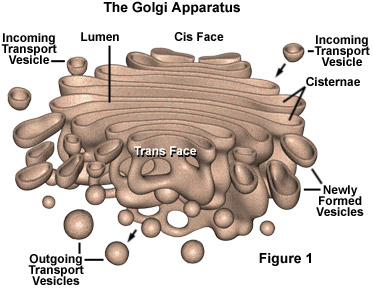
The Golgi apparatus (GA), also called Golgi body or Golgi complex and found universally in both plant and animal cells, is typically comprised of a series of five to eight cup-shaped, membrane-covered sacs called cisternae that look something like a stack of deflated balloons. In some unicellular flagellates, however, as many as 60 cisternae may combine to make up the Golgi apparatus. Similarly, the number of Golgi bodies in a cell varies according to its function. Animal cells generally contain between ten and twenty Golgi stacks per cell, which are linked into a single complex by tubular connections between cisternae. This complex is usually located close to the cell nucleus.
Due to its relatively large size, the Golgi apparatus was one of the first organelles ever observed. In 1897, an Italian physician named Camillo Golgi, who was investigating the nervous system by using a new staining technique he developed (and which is still sometimes used today; known as Golgi staining or Golgi impregnation), observed in a sample under his light microscope a cellular structure that he termed the internal reticular apparatus. Soon after he publicly announced his discovery in 1898, the structure was named after him, becoming universally known as the Golgi apparatus. Yet, many scientists did not believe that what Golgi observed was a real organelle present in the cell and instead argued that the apparent body was a visual distortion caused by staining. The invention of the electron microscope in the twentieth century finally confirmed that the Golgi apparatus is a cellular organelle.
The Golgi apparatus is often considered the distribution and shipping department for the cell's chemical products. It modifies proteins and lipids (fats) that have been built in the endoplasmic reticulum and prepares them for export outside of the cell or for transport to other locations in the cell. Proteins and lipids built in the smooth and rough endoplasmic reticulum bud off in tiny bubble-like vesicles that move through the cytoplasm until they reach the Golgi complex. The vesicles fuse with the Golgi membranes and release their internally stored molecules into the organelle. Once inside, the compounds are further processed by the Golgi apparatus, which adds molecules or chops tiny pieces off the ends. When completed, the product is extruded from the GA in a vesicle and directed to its final destination inside or outside the cell. The exported products are secretions of proteins or glycoproteins that are part of the cell's function in the organism. Other products are returned to the endoplasmic reticulum or may undergo maturation to become lysosomes.
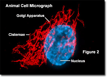
The modifications to molecules that take place in the Golgi apparatus occur in an orderly fashion. Each Golgi stack has two distinct ends, or faces. The cis face of a Golgi stack is the end of the organelle where substances enter from the endoplasmic reticulum for processing, while the trans face is where they exit in the form of smaller detached vesicles. Consequently, the cis face is found near the endoplasmic reticulum, from whence most of the material it receives comes, and the trans face is positioned near the plasma membrane of the cell, to where many of the substances it modifies are shipped. The chemical make-up of each face is different and the enzymes contained in the lumens (inner open spaces) of the cisternae between the faces are distinctive. Illustrated in Figure 2 is a fluorescence digital image taken through a microscope of the Golgi apparatus (pseudocolored red) in a typical animal cell. Note the close proximity of the Golgi membranes to the cell nucleus.
Proteins, carbohydrates, phospholipids, and other molecules formed in the endoplasmic reticulum are transported to the Golgi apparatus to be biochemically modified during their transition from the cis to the trans poles of the complex. Enzymes present in the Golgi lumen modify the carbohydrate (or sugar) portion of glycoproteins by adding or subtracting individual sugar monomers. In addition, the Golgi apparatus manufactures a variety of macromolecules on its own, including a variety of polysaccharides. The Golgi complex in plant cells produces pectins and other polysaccharides specifically needed by for plant structure and metabolism. The products exported by the Golgi apparatus through the trans face eventually fuse with the plasma membrane of the cell. Among the most important duties of the Golgi apparatus is to sort the wide variety of macromolecules produced by the cell and target them for distribution to their proper location. Specialized molecular identification labels or tags, such as phosphate groups, are added by the Golgi enzymes to aid in this sorting effort.