
Cilia and Flagella
Cilia and flagella are motile cellular appendages found in most microorganisms and animals, but not in higher plants. In multicellular organisms, cilia function to move a cell or group of cells or to help transport fluid or materials past them. The respiratory tract in humans is lined with cilia that keep inhaled dust, smog, and potentially harmful microorganisms from entering the lungs. Among other tasks, cilia also generate water currents to carry food and oxygen past the gills of clams and transport food through the digestive systems of snails. Flagella are found primarily on gametes, but create the water currents necessary for respiration and circulation in sponges and coelenterates as well. For single-celled eukaryotes, cilia and flagella are essential for the locomotion of individual organisms. Protozoans belonging to the phylum Ciliophora are covered with cilia, while flagella are a characteristic of the protozoan group Mastigophora.
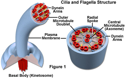
In eukaryotic cells, cilia and flagella contain the motor protein dynein and microtubules, which are composed of linear polymers of globular proteins called tubulin. The core of each of the structures is termed the axoneme and contains two central microtubules that are surrounded by an outer ring of nine doublet microtubules. One full microtubule and one partial microtubule, the latter of which shares a tubule wall with the other microtubule, comprise each doublet microtubule (see Figure 1). Dynein molecules are located around the circumference of the axoneme at regular intervals along its length where they bridge the gaps between adjacent microtubule doublets.
A plasma membrane surrounds the entire axoneme complex, which is attached to the cell at a structure termed the basal body (also known as a kinetosome). Basal bodies maintain the basic outer ring structure of the axoneme, but each of the nine sets of circumferential filaments is composed of three microtubules, rather than a doublet of microtubules. Thus, the basal body is structurally identical to the centrioles that are found in the centrosome located near the nucleus of the cell. In some organisms, such as the unicellular Chlamydomonas, basal bodies are locationally and functionally altered into centrioles and their flagella resorbed before cell division.
Eukaryotic cilia and flagella are generally differentiated based on size and number: cilia are usually shorter and occur together in much greater numbers than flagella, which are often solitary. The structures also exhibit somewhat different types of motion, though in both cases movement is generated by the activation of dynein and the resultant bending of the axoneme. The movement of cilia is often described as whip-like, or compared to the breast stroke in swimming. Adjacent cilia move almost simultaneously (but not quite), so that in groups of cilia, wave-like patterns of motion occur. Flagella, however, exhibit a smooth, independent undulatory type of movement in eukaryotes. Prokaryotic flagella, which have a completely different structure built from the protein flagellin, move in a rotating fashion powered by the basal motor.
Defects in the cilia and flagella of human cells are associated with some notable medical problems. For example, a hereditary condition known as Kartagener's syndrome is caused by problems with the dynein arms that extend between the microtubules present in the axoneme, and is characterized by recurrent respiratory infections related to the inability of cilia in the respiratory tract to clear away bacteria or other materials. The disease also results in male sterility due to the inability of sperm cells to propel themselves via flagella. Damage to respiratory cilia may also be acquired rather than inherited and is most commonly linked to smoking cigarettes. Bronchitis, for instance, is often triggered by a build-up of mucus and tar in the lungs that cannot be properly removed due to smoking-related impairment of cilia.