
Centrioles
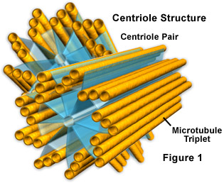
Found only in animal cells, these paired organelles are typically located together near the nucleus in the centrosome, a granular mass that serves as an organizing center for microtubules. Within the centrosome, the centrioles are positioned so that they are at right angles to each other, as illustrated in Figure 1. Each centriole is made of nine bundles of microtubules (three per bundle) arranged in a ring.
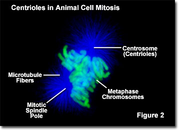
Centrioles play a notable role in cell division. During interphase of an animal cell, the centrioles and other components of the centrosome are duplicated, though scientists are not yet sure how this duplication takes place. At first the two pairs of centrioles remain in close proximity to each other, but as mitosis initiates, the original centrosome divides and the pairs are split up so that one set of centrioles is located in each of the new microtubule-organizing centers. These new centers radiate microtubules in star-shaped clusters known as asters. As the asters move to opposing poles of the cells, the microtubules, with the help of the centrioles, become organized into a spindle-shaped formation that spans the cell (see Figure 2). These spindle fibers act as guides for the alignment of the chromosomes as they separate later during the process of cell division.
Though centrioles play a role in the mitosis of animal cells, plant cells are able to reproduce without them. Researchers have, therefore, been very interested in determining exactly how important the organelles really are. Studies have shown that certain animal cells, particularly female gametes (oocytes), can successfully divide even when their centrioles are destroyed. Some investigators have also found, however, that the absence of centrioles in animal cells is associated with an increased number of divisional errors and substantial delays in the mitotic process, especially before chromosome segregation. Consequently, it has been suggested that centrioles evolved as a refinement of the cell, making mitosis a much more efficient and less error-prone process.
In cells that feature cilia or flagella, basal bodies, which exhibit the same structural form as centrioles, are present. These assemblies are located, however, near the cell surface at the base of each cilium or flagellum, rather than in the centrosome near the nucleus. Basal bodies are anchored in their cytoplasmic locations by what is called a rootlet system in the cell. In some organisms, such as the unicellular Chlamydomonas, basal bodies change their location and are functionally converted to centrioles before the mitotic process.