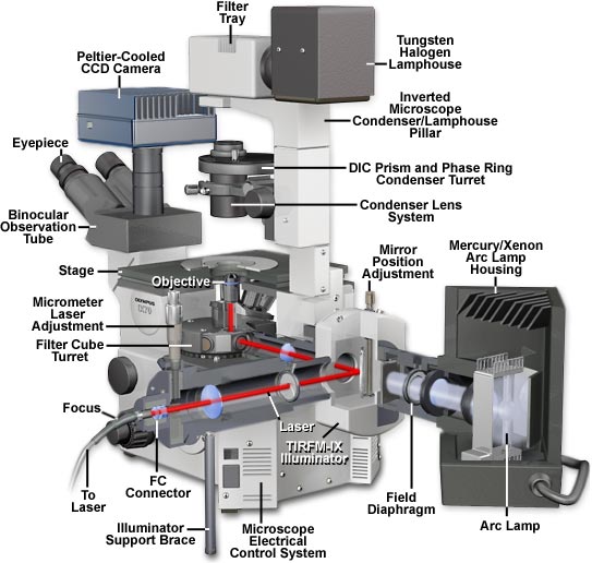
Olympus IX70 Inverted Microscope Cutaway Diagram
|
The Olympus IX70 inverted tissue culture microscope is a research-level instrument capable of imaging specimens in a variety of illumination modes including brightfield, darkfield, phase contrast, Hoffman modulation contrast, fluorescence, and differential interference contrast. A recent accessory for this popular microscope is the TIRFM-IX illuminator, which attaches to the rear lamphouse port and contains an FC connector for an external laser source and a port for a mercury or xenon lamp housing. A view of the microscope from the rear (illustrated above) reveals how the total internal reflection illuminator appears when attached to the instrument. The central feature of the TIRFM-IX attachment is a dual port intermediate tube (the UD-P port) that was originally designed to mount two cameras onto a single microscope port. Light is directed to the individual camera tubes (or illumination sources) with BX and BX2 series mirror units (fluorescence cubes), which also serve to restrict wavelength passage with interference filters. For simplicity and ease of illustration, a swivel-joint mirror assembly is substituted for the mirror block in the diagram presented above. Attached to the UD-P intermediate piece are three ports: one for the microscope illuminator, a second for the laser FC connector, and a third port that houses a field diaphragm and shutter and contains a connector for a mercury or xenon arc lamphouse. The laser tube portion of the illuminator also contains a shutter and field diaphragm, as well as a centering mechanism for the laser beam. Adjustment of the laser light source for TIRFM is accomplished by means of a micrometer that translates the position of beam entry into the objective rear aperture. Illumination into the microscope is controlled by a lever housed in the UD-P unit that engages the mirror block to position it into the light pathway for simultaneous arc lamp and laser excitation. Removal of the mirror block, by lifting the control lever, results in exclusive illumination from the arc lamp source. Once light (either arc or laser) has entered the microscope rear port, it is directed through the optical train in a traditional manner. Contributing Authors Mortimer Abramowitz and William K. Fester - Olympus America, Inc., Two Corporate Center Drive., Melville, New York, 11747. Michael W. Davidson - National High Magnetic Field Laboratory, 1800 East Paul Dirac Dr., The Florida State University, Tallahassee, Florida, 32310. |
© 1995-2025 by Michael W. Davidson and The Florida State University. All Rights Reserved. No images, graphics, software, scripts, or applets may be reproduced or used in any manner without permission from the copyright holders. Use of this website means you agree to all of the Legal Terms and Conditions set forth by the owners.
This website is maintained by our
|
