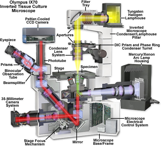
Olympus IX70 Inverted Microscope Cutaway Diagram
|
Microscopes with an inverted-style frame are designed primarily for tissue culture applications and are capable of producing fluorescence illumination through an episcopic and optical pathway. Epi-illuminators usually consist of a mercury or xenon lamphouse (or laser system) stationed in a port at the rear of the microscope frame. Fluorescence illumination from the arc lamp passes through a collector lens and into a cube that contains a set of interference filters, including a dichroic mirror, barrier filter, and excitation filter. Light reflected from the dichroic mirror is restricted in wavelength by the excitation filter and enters the objective (now acting as a condenser) to bathe the specimen with a cone of illumination whose size and shape is determined by the objective numerical aperture. Secondary fluorescence, emitted by the specimen, returns through the objective, dichroic mirror and barrier filter before being routed through the microscope optical train. The microscope presented above contains a trinocular observation tube that is equipped with a port and extension tube for mounting a traditional or CCD camera system (a Peltier-cooled CCD camera is illustrated). Another port, located near the base at the front of the microscope, can also serve as an attachment point for a camera system (a traditional 35-millimeter camera is shown in the figure). In the figure presented above, wide-spectrum fluorescence illumination is filtered to produce a narrow bandwidth of green excitation wavelengths, which are capable of exciting specific fluorophores in the specimen. Secondary fluorescence (red light) passes back through the objective and is distributed throughout the microscope optical system. Transmitted illumination is provided by a tungsten-halogen lamphouse that is positioned on the microscope pillar, above the stage. Light from the lamphouse passes through a collector lens, a series of filters, and the field diaphragm before entering the condenser front aperture. After being focused by the condenser lens elements, transmitted illumination is projected onto the specimen, which is placed on the stage. The light that is diffracted, refracted, and not absorbed by the specimen continues through the objective and into the microscope optical train where it can be directed to the eyepieces or to a camera system. Contributing Authors William K. Fester and Mortimer Abramowitz - Olympus America, Inc., Two Corporate Center Drive., Melville, New York, 11747. Michael W. Davidson - National High Magnetic Field Laboratory, 1800 East Paul Dirac Dr., The Florida State University, Tallahassee, Florida, 32310. |
© 1995-2025 by Michael W. Davidson and The Florida State University. All Rights Reserved. No images, graphics, software, scripts, or applets may be reproduced or used in any manner without permission from the copyright holders. Use of this website means you agree to all of the Legal Terms and Conditions set forth by the owners.
This website is maintained by our
|