|
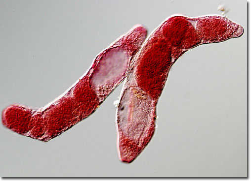
F. buski was first described in 1843 by English surgeon George Busk, who found the parasite in the duodenum of a sailor. The complex lifecycle of the trematode was not understood, however, until Claude H. Barlow’s determination of it in 1925. As he revealed, immature eggs of F. buski are discharged from humans or other definitive hosts with bodily wastes. If the eggs then reach water, they hatch in 3 to 7 weeks and form miracidia, organisms that looks similar to ciliated protozoa. The miracidia penetrate the soft tissues of snails and develop into a sporocyst state. The sporocysts then produce many daughter stages called rediae that asexually reproduce to yield cercariae. The cercariae, which feature a tail-like structure that enables them to swim, exit the snail host and encyst on nearby vegetation, where they develop into metacercariae.
|
