Differential Interference Contrast Image Gallery
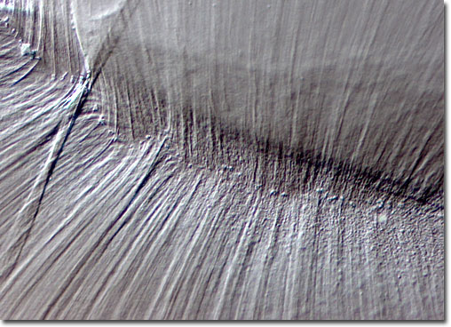
Chicken Embryo Lens
|
Although initially all cells within a chicken embryo look alike, they quickly begin to develop into specialized structures. Within the first 24 hours of embryonic development, the head becomes distinguishable, the foregut forms, blood begins to accumulate in areas that will become parts of the vascular system, and the eye starts to take shape. Developmental progression moves swiftly, and on the second day the lenses of the eyes materialize, the vascular system takes a definite form, and the heart begins beating. The lightning-fast pace of growth and change continues, each feature developing within a set period of time, until a few short weeks later a full-formed and functional baby chicken, complete with claws and feathers, taps it way out of its shell to first see the light of day. |
© 1995-2025 by Michael W. Davidson and The Florida State University. All Rights Reserved. No images, graphics, software, scripts, or applets may be reproduced or used in any manner without permission from the copyright holders. Use of this website means you agree to all of the Legal Terms and Conditions set forth by the owners.
This website is maintained by our
|