Brightfield Microscopy Digital Image Gallery
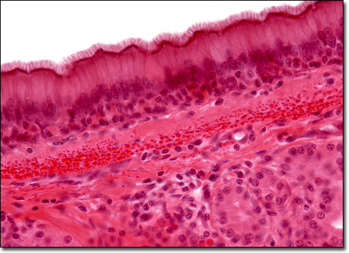
Pseudostratified Ciliated Columnar Epithelium
|
Unlike the epithelium of the skin, a pseudostratified ciliated columnar epithelium appears to have multiple layers, but is actually only comprised of a single sheet of cells. The positioning of the nuclei within the individual columnar cells causes this illusion. These structures, which are easily identifiable with the help of a microscope, are found at various levels, creating a stratified appearance. A microscope also facilitates the observation of the tiny hairlike cilia that line the cells. Found most heavily along the respiratory tract, pseudostratified ciliated columnar epithelial cells help trap and transport particles brought in through the nasal passages and lungs. |
© 1995-2025 by Michael W. Davidson and The Florida State University. All Rights Reserved. No images, graphics, software, scripts, or applets may be reproduced or used in any manner without permission from the copyright holders. Use of this website means you agree to all of the Legal Terms and Conditions set forth by the owners.
This website is maintained by our
|