Brightfield Microscopy Digital Image Gallery
Ductus Deferens
|
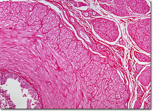
Thick-walled, the ductus deferens is composed of several layers of tissue. Then innermost layer is a folded mucous membrane that is always moist. Encircling this layer are three layers of muscle tissue, which provide the tube with the ability to contract, a necessary action for the conveyance of bodily fluids and sperm. As the ductus deferens rises upwards from the epididymis towards the bladder, it also comes to be surrounded by nerve fibers, arteries, and veins, and the complex structure further encased in connective tissue is referred to as the spermatic cord. When the tube reaches the level of the bladder, however, it becomes separated from the connective tissue and extends across the top of the bladder and turns downward near the back of the structure, ending in an enlarged ampulla, which both stores and adds secretions to semen. |
© 1995-2025 by Michael W. Davidson and The Florida State University. All Rights Reserved. No images, graphics, software, scripts, or applets may be reproduced or used in any manner without permission from the copyright holders. Use of this website means you agree to all of the Legal Terms and Conditions set forth by the owners.
This website is maintained by our
|