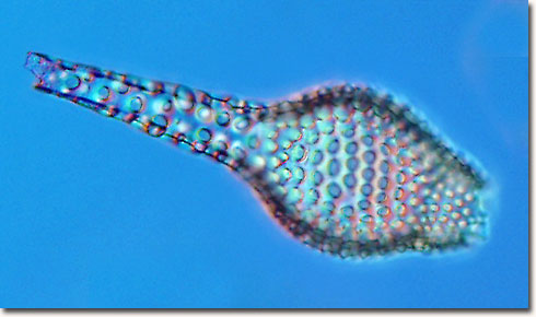
Radiolarian in Differential Interference Contrast (DIC)
|
The radiolarian skeleton (test) presented above was captured utilizing an Olympus BX60 optical microscope operating in differential interference contrast (DIC) mode with a 20x plan fluorite objective. Successive optical serial sections of the specimen were recorded on a Nikon DXM 1200 camera system attached to the microscope, and merged together with an image-editing software package (Adobe Photoshop). By adding optical sections and removing out-of-focus blur, a more detailed image of the skeleton can be obtained. |
© 1995-2025 by Michael W. Davidson and The Florida State University. All Rights Reserved. No images, graphics, software, scripts, or applets may be reproduced or used in any manner without permission from the copyright holders. Use of this website means you agree to all of the Legal Terms and Conditions set forth by the owners.
This website is maintained by our
|