Crystals Within Haversian Canals
|
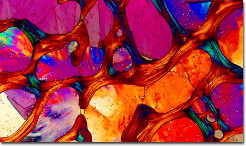
The osteon, which is comprised of many layers of material, is the principal structural unit of mature compact bone. Cylindrical in shape, osteons are readily seen in cross sections of bone, appearing as clusters of concentric circles, each of which surrounds a central canal. Named Haversian canals in honor of Clopton Havers, the seventeenth-century English anatomist and physician who discovered them, these important formations house diminutive blood vessels and nerves in living organisms, though they appear filled with crystals in the photomicrograph of fossilized dinosaur bone shown above. The primary purpose of the vessels typically found in Haversian canals is to provide a blood supply to bone cells, which are also known as osteocytes. |
© 1995-2025 by Michael W. Davidson and The Florida State University. All Rights Reserved. No images, graphics, software, scripts, or applets may be reproduced or used in any manner without permission from the copyright holders. Use of this website means you agree to all of the Legal Terms and Conditions set forth by the owners.
This website is maintained by our
|