Starch Granules in Potato Tissue
|
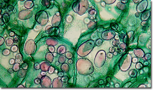
Presented in the digital image above is a thin section photomicrograph of potato (Solanum tuberosum) tissue illustrating a portion of mature tuber with periderm, cortex, reduced vascular tissues, and starch storage granules. The section was stained with a quadruple mixture containing the following ingredients: safranin O (stains nuclei, chromosomes, lignified and cutinized cell walls red); fast green (stains cytoplasm and cellulose cell walls green); crystal violet (stains starch grains purple as evident in the photomicrograph); and orange G (stains acidophilic cytoplasm and cell walls yellow to green). Digital images were captured in brightfield illumination with a Nikon Eclipse E600 microscope attached to a DXM 1200 digital camera system. Note the large number of significant starch granules present in this tissue. |
© 1995-2025 by Michael W. Davidson and The Florida State University. All Rights Reserved. No images, graphics, software, scripts, or applets may be reproduced or used in any manner without permission from the copyright holders. Use of this website means you agree to all of the Legal Terms and Conditions set forth by the owners.
This website is maintained by our
|