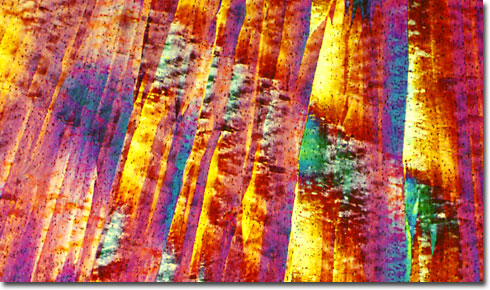
Crystallized Cheese Proteins
|
Presented in the digital image above is a polarized light microscope image of cheese proteins that were extracted and recrystallized to produce a kaleidoscopic array of color. The birefringence of this specimen was captured utilizing polarized light and a 540-nanometer retardation plate with a strain-free 10x objective on a Nikon Eclipse E600 microscope. Processed cheese was shredded, placed into a container with 10N hydrochloric acid and heated at 100 degrees Celsius for 12 hours. The resulting slurry was extracted with methylene chloride and the residue re-dissolved in acetone and filtered. Small aliquots of acetone were sandwiched between a microscope slide and coverslip and allowed to slowly evaporate to create the crystal formations illustrated in the digital image. The exact nature and composition of the substances recovered from the acetone solution are unknown. |
© 1995-2025 by Michael W. Davidson and The Florida State University. All Rights Reserved. No images, graphics, software, scripts, or applets may be reproduced or used in any manner without permission from the copyright holders. Use of this website means you agree to all of the Legal Terms and Conditions set forth by the owners.
This website is maintained by our
|
