The Brain
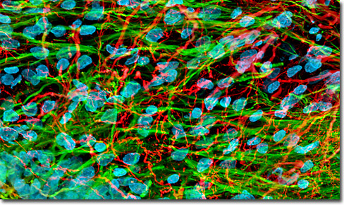
Rat Brain Hippocampus Neurons
|
Throughout history humans have sought to understand the origin of their own consciousness and have inquiringly searched for answers to the innumerable questions such a quest inevitably raises regarding the generation of thought, the formation of memories, the basis of emotions, and similar issues. Philosophers, theologians, poets, and early scientists have all held varying views. Aristotle, for instance, alleged that intelligence resided in the heart, and his influence on Western thought was so strong that nearly 2,000 years later William Shakespeare wrote, “Go to your bosom; Knock there, and ask your heart what it doth know.” Modern science attests, however, that is not the business of the heart to “know” anything, since the awareness and understanding that constitutes knowledge is seated in an entirely different organ—the brain. The human brain is a remarkable example of evolution. Paleontological evidence suggests that the organ has undergone significant changes in both its overall size and the relative size of its component parts. The physical changes have apparently been accompanied by important modifications in the mental processes, humans having become increasingly capable of more and more complex activities over time. The phenomenon of evolution is similarly evident among the diverse nervous systems that belong to the members of the animal kingdom in general. Even the simplest, most primitive prokaryotes are able to sense and respond to certain environmental cues, such as movement toward or away from a chemical stimulant (chemotaxis), but only more advanced organisms possess the networks of nerve cells necessary to better perceive the environment and carry out more complex responses based on that perception. 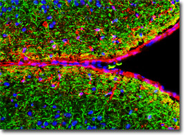
Cerebral Cortex Studies of the embryonic development of the vertebrate brain suggest that the progression parallels the evolution of the organ. The brain and the spinal cord, which comprise the vertebrate central nervous system, develop from the tube-like dorsal nerve cord of the embryo. In the early stages of nerve cord differentiation, three anterior bulges, known as the forebrain, midbrain, and hindbrain are observable. These three sections represent the earliest evolutionary structures considered to comprise a brain. Later in evolutionary history, additional structural divisions appeared, as they do further along in embryonic development. By the time a human embryo is five weeks old, the forebrain has enlarged and differentiated into two sections, called the telencephalon and the diencephalon, and the hindbrain has differentiated into the metencephelon and myelencephalon. The midbrain is also modified, though it is still only considered to comprise a single structure, the mesencephalon. By the time the human brain has completed its development, the telencephalon has given rise to the cerebrum, which is considered the organ’s most highly evolved structure. In advanced vertebrates, including humans, the cerebrum is so large that it covers almost all other structures of the brain. The fully developed human brain also features several other regions that have differentiated from structures present in earlier developmental stages. In the adult, the region of the brain known before only as the diencephalon is differentiated into three distinguishable centers called the thalamus, hypothalamus, and the epithalamus, the metencephelon is differentiated into the pons and cerebellum, the myelencephalon has given rise to the medulla oblongata, and the mesencephalon has changed into what is called midbrain, though it is much more advanced than the midbrain present early in embryonic development. Several structures of the adult human brain are located deep within the organ and appear to form a stem that the cap-like cerebellum sits upon. Collectively the structures, the medulla oblongata, the pons, and the midbrain, are called the brainstem. The brainstem is what connects the brain to the anterior region of the spinal cord and serves as the main communication pathway between other regions of the brain and the spinal cord and peripheral nervous system. The brain stem also controls several functions, primarily those that involve essential, automatic activities, such as respiration, the beating of the heart, and homeostasis. Within the core of the brainstem is the reticular formation, a nerve cell system that functions in filtering out familiar or habitual input. A part of this system, called the reticular activating system, is heavily involved in the regulation of sleep and states of arousal. A more detailed understanding of the brainstem can be gained by considering the component parts of the structure individually. 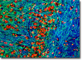
Coronal Hippocampus Thin Section The medulla oblongata, often referred to simply as the medulla, is the lowermost portion of the vertebrate brain and is directly continuous with the spinal cord. At its other end, the medulla is linked to the pons. The medulla encloses one of the four ventricles of the brain, which are cavities filled with cerebrospinal fluid that are linked to the central canal of the spinal cord. Two types of nervous tissue, commonly called gray matter and white matter, comprise the medulla, as well as the other parts of the brain. Characteristically gray matter predominantly consists of nerve cell bodies whose nuclei lend the tissue a gray appearance in section, whereas white matter is composed mainly of myelinated axons, its name stemming from the pearly white appearance of the insulating myelin. Due to the different makeup of gray and white matter, the tissue types function in distinct ways, with gray matter chiefly being involved in processing information and white matter in communicating information. Unlike most other parts of the brain, the white matter of the medulla is exterior to its gray matter, which surrounds the ventricle. Functionally the medulla is extremely important in an integrative capacity, but it also contains nerve centers that regulate various involuntary nervous activities, including digestion, heart activity, vomiting, swallowing, sleeping, and breathing. Coordination of large-scale movement is another key function of the medulla. Notably, as most descending axons that send signals regarding movement pass through the medulla to the spinal cord, they cross over to the opposite side of the central nervous system from which they originated. Accordingly, the left side of the brain usually directs the movement of the right side of the body, and the right side of the organ controls most of the movement of the left side of the body. In addition to descending axons, seven of the 12 sets of cranial nerves extend from the medulla. The pons exhibits a horseshoe-like shape and is located directly above the medulla and below the midbrain. The structure functions in the transmission of information between higher regions of the brain and the spinal cord and between the cerebrum and the cerebellum. Nervous centers that assist in the control of a number of autonomic activities, such as breathing, sleep, and arousal, are also found in the pons. Four pairs of cranial nerves originate in the pons. 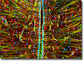
Intermediate Filaments in Brain Tissue The final section of the brainstem, the midbrain, plays a major part in the integration of sensory input in many vertebrates, but in humans and other mammals, its role is somewhat reduced, functioning primarily as a communication corridor to the cerebral hemispheres. In nonmammalian vertebrates, for instance, a pair of specialized centers called the superior colliculi are very prominent and sometimes serve as the sole location where visual information may be integrated, but in mammals visual integration takes place in the cerebrum. Coordination of visual reflexes is the primary role of mammalian superior colliculi. Another pair of specialized centers in the midbrain, known as the inferior colliculi, are a central part of the auditory system. Located posterior to the brain stem and connected to it via bundled nerves is the cerebellum, the moniker of which was coined to reflect its appearance, which is similar to that of a small brain (cerebellum is the diminutive form of the Latin word for brain). The cerebellum is relatively large in humans and is divided into two lateral hemispheres, similar to the cerebrum. An outer cortex of gray matter and an inner region of white matter comprise the hemispheres, each of which is subdivided into three lobes. Different types of input are received by each of the lobes: the flocculonodular lobe collects information from the inner ear vestibule (a balance, rather than auditory, component), the anterior lobe receives impulses from the spinal cord, and the posterior lobe communicates with the cerebrum. All of the input received by the various lobes is integrated in the cortex of the cerebellum. The coordinated activity of the multiple parts of the cerebellum enables this region of the brain to control refined, coordinated muscle movements and balance. Similar to the brainstem, three different regions comprise the diencephalon of the adult human brain: the epithalamus, the thalamus, and the hypothalamus. Within the epithalamus is a group of capillaries collectively called a choroid plexus that manufactures cerebrospinal fluid. The epithalamus is also the site of the pineal gland, a constituent of the endocrine system that secretes the hormone melatonin, which appears to play an important role in regulating the sleep cycle and other cyclical behaviors. In mammals, the pineal gland is positioned near the center of the brain, but in some lower vertebrates the gland is located in closer proximity to the surface of the brain. Modern studies suggest that the pineal gland was possibly an evolutionary predecessor of the eye, a far cry from the belief of the seventeenth century philosopher René Descartes, who suggested it accommodated the soul. 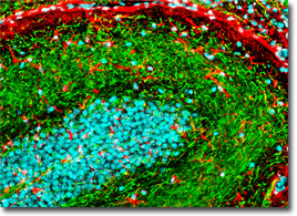
Mouse Brain Post Hypothalamus Region The hypothalamus is also associated with the rhythmic behaviors of the body, especially the daily rhythms known as circadian rhythms. The suprachiasmatic nuclei located in the hypothalamus are thought to serve as one of the body’s primary biological clocks, and the paired structures are linked to the pineal gland so that they function in the regulation of melatonin secretion. The hypothalamus also interacts with the pituitary gland and heavily influences its activity, including the release of growth hormone and luteinizing hormone, among others. Weight, body heat, hunger, thirst, fluid intake, pleasure, and mating behavior are some of the many other properties and activities heavily impacted by the hypothalamus. Unlike the other regions of the brain that comprise the diencephalon, the thalamus does not appear to play a notable role in cyclical behaviors. Instead, the double-lobed mass of gray matter that is located superior to the hypothalamus functions in motor control by being the primary recipient of motor information transmitted by the cerebrum. The thalamus is also a receiver of all auditory, somatosensory, and visual sensory signals from diverse regions of the brain, serving as the final gateway impulses must pass on their way to the cerebrum. Within the skull, the cerebrum occupies the uppermost region. The two hemispheres of the cerebrum are separated by a deep groove called the longitudinal cerebral fissure, and each of the sections is divided into an inner core of white matter and an outer layer of gray matter known as the cerebral cortex. Located deep inside of the white matter are clustered nuclei termed basal nuclei that appear to be primarily concerned with the planning of sequences of movement and learning to perform them again. The site of most activity in the cerebrum, however, is the cerebral cortex. In mammals, the cerebral cortex is large and complex, while only the most basic components of this region of the brain are present in reptiles. 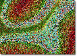
Cerebellum The neocortex, an outer cortical stratum composed of six layers of neurons, is only found in mammalian species, and is most highly developed in humans. Some types of brain activity are allied with a single layer of the cerebral cortex, but many processes are believed to be interactively regulated. Advanced behavior and cognitive capacity is associated with the relative size of the neocortex and the extent to which the region is convoluted. The human neocortex is less than 5 millimeters thick, but due to its extensive wrinkling has a surface area of approximately 0.5 meters square. Next to humans, nonhuman primates, porpoises, and whales possess some of the most highly developed neocortices. In addition to the neocortex, the cortical region of the human brain contains more primitive components called the olfactory cortex and the hippocampus that occur in reptiles as well as mammals. The mammalian versions of these structures, however, are associated with other regions of the cortex, hypothalamus, and thalamus in a ring-like assembly centered around the brainstem known as the limbic system. The emotional responses or feelings that mammals experience are produced by the limbic system, which closely interacts with other parts of the brain. The limbic system also is a functional center for long-term memory. Each hemisphere of the cerebrum is traditionally considered to be comprised of four outwardly observable lobes: the frontal lobe, temporal lobe, occipital lobe, and parietal lobe. A fifth lobe, called the insular lobe, is situated beneath them within a fissure that separates the parietal and temporal lobes. Various regions of the lobes are specialized for certain functions, such as receiving specific types of sensory information (primary sensory areas) or integrating sensory information with input received from other regions of the brain (association areas). For example, the somatosensory cortex located in the parietal lobe receives input acquired from pain, temperature, and other receptors in the body, while the auditory association area in the temporal lobe integrates and processes auditory information. In humans, association areas located in the frontal lobes usually only receive signals that have already passed through other association areas. This additional step of association is thought to be strongly related to the human capacity for advanced mental activity. Without evolving such cognitive ability, humans, who must compete with countless other species for survival, may not have been so successful at inhabiting the Earth, and most certainly would not be pondering issues involving the structure and function of the brain. Despite the incredible mental potential that characterizes humanity, however, scientists appear to have only begun to answer many of the questions that have been raised about the brain. And, as should be expected considering the astoundingly inquisitive nature of the organ that dictates almost all human activity, as more information is learned, more questions come to mind. |
© 1995-2025 by Michael W. Davidson and The Florida State University. All Rights Reserved. No images, graphics, software, scripts, or applets may be reproduced or used in any manner without permission from the copyright holders. Use of this website means you agree to all of the Legal Terms and Conditions set forth by the owners.
This website is maintained by our
|