High Density Liquid Crystalline DNA
|
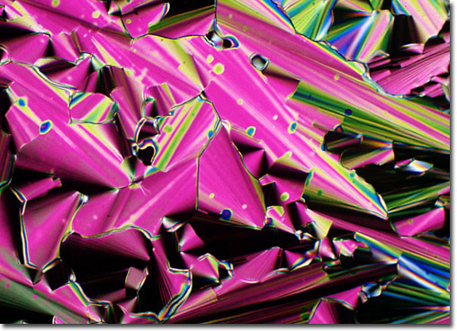
The focal conic liquid crystalline texture shows both a Schlieren pattern surrounding the edges of the cones and a color transition from pinkish-purple to green. Differences in thickness of the DNA solution sandwiched between the cover glass and microscope slide are responsible for the color shift. Spots on the conical textures are the result of miniature gas bubbles in the DNA solution. The DNA concentration for this specimen is approximately 450 milligrams per millimeter, and the magnification is approximately 320x. The digital image was recorded on Fujichrome 64T transparency film using a Nikon Optiphot-Pol microscope with crossed polarized illumination. Exposures were recorded about 2.5 f-steps under the recommended value given by an in-camera photomultiplier and were push-processed about 1.25 f-steps in the first E-6 developer. |
© 1995-2025 by Michael W. Davidson and The Florida State University. All Rights Reserved. No images, graphics, software, scripts, or applets may be reproduced or used in any manner without permission from the copyright holders. Use of this website means you agree to all of the Legal Terms and Conditions set forth by the owners.
This website is maintained by our
|