High Density Liquid Crystalline DNA
|
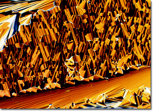
The vast size difference in focal conic texture size exhibited by the DNA sample presented above indicates a variety of crystallization rates. The smaller textures in the top portion of the micrograph formed at a much faster rate than the larger texture at the bottom. The DNA concentration for this specimen is approximately 450 milligrams per millimeter, and the magnification is approximately 350x. The digital image presented above was originally recorded on Fujichrome 64T transparency film using a Nikon Optiphot-Pol microscope with crossed polarized illumination. Exposures were recorded about 2.5 f-steps under the recommended value given by an in-camera photomultiplier and were push-processed approximately 1.5 f-steps in the first E-6 developer. |
© 1995-2025 by Michael W. Davidson and The Florida State University. All Rights Reserved. No images, graphics, software, scripts, or applets may be reproduced or used in any manner without permission from the copyright holders. Use of this website means you agree to all of the Legal Terms and Conditions set forth by the owners.
This website is maintained by our
|