Is That Shirt Ascorbic Acid?
National High Magnetic Field Laboratory
The Florida State University
Tallahassee, Florida 32306
As any three-year-old will show you, wearing food is more fun than eating it. That was not quite what Michael W. Davidson, a biophysicist from the National High Magnetic Field Laboratory at Florida State University, had in mind in the mid-1980s. Back then he was using photomicrography to see how DNA crystals organize themselves in such places as sperm heads and bacteria.
Now Davidson finds his colorful and unusual images-of medicines, hormones, superconductors and pesticides, to name a few-the focus of a mini-industry. Vitamins and other images from Davidson's laboratory will be smeared on T-shirts, neckties and other body wear and will be available this summer-without the laundry bill. The scientific images have already begun to work their way onto other mass-market items, among them greeting cards, gift-wrapping and even a New Age album cover.
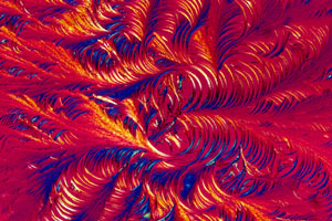
The graphics are the result of improved film emulsions and camera technology coupled to various kinds of microscope techniques. The primary technique Davidson relies on is optically polarized light micrography, in which, he explains, "the polarized light goes through, and the crystals bend the light." The degree of refraction and hence the resulting colors depend on the molecular orientation and the thickness of the crystal. The method is ideal for many biological molecules, including the AIDS drug zidovudine (AZT) (below).
|
|
AIDS drug zidovudine (AZT) |
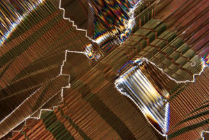
For samples too thick to transmit light, Davidson uses the reflected differential interference contrast method. This technique calls for the apparatus to split the illumination into two components. One beam bounces off the sample and then recombines with the other. The beams interfere to produce, with the appropriate filters and prisms, a colorful, three-dimensional effect, such as that of the high-temperature superconductor yttrium-barium-copper oxide silk-screened onto an alumina substrate (below).
Davidson can now use his imaging technique to create surreal artwork he calls microscapes. For instance, his "Nuclear Sunrise" photograph (below) is made from multiple exposures of different compounds. The desert foreground is ascorbic acid, the morning sky is stretched polyethylene and the stars (mostly obscured by the polyethylene) are small spheres of polybenzyl-L-glutamate. The rising sun is an image of the microscope diaphragm, slightly out of focus. Two of his microscapes have won Davidson grand prizes in photomicroscopy competitions sponsored by Polaroid, and New Age musician Richard Burmer is using one for the cover of an upcoming album.
Artistic recognition rarely translates into untold wealth, but it helps with the laboratory overhead. `About a year ago we decided to make some money on things that aren't ordinarily funded," says Davidson, who estimates that he sent out portfolios to more than 300 companies. His shotgun approach paid off. Amber Lotus, a paper products company in Oakland, Calif., already markets calendars, gift wrap and greeting cards with Davidson's motifs. In late July, Danskin, an apparel firm based in New York City, expects to introduce T-shirts and exercise wear-featuring images of vitamins. Davidson has signed licensing agreements with several other firms as well. Florida State gets the proceeds, so "the people in the laboratory are really happy," Davidson notes. "We never expected it to get to this point."