SOME ARTISTIC TECHNIQUES IN PHOTOMICROGRAPHY
Michael W. Davidson
The National High Magnetic Field Laboratory (NHFML)
and the Supercomputer Computations
Research Institute (SCRI)
The Florida State University
Tallahassee, Florida 32306
Telephone: 850-644-6114 Telefax: 850-644-8281
INTRODUCTION
The inherent beauty of science has recently become more accessible to the general public due to an escalation in the publication of multi-color computer-generated graphics images as well as photomicrographs and other images of scientific interest. These images are appearing on the covers of many scientific and trade periodicals (currently numbering over 250) where they serve as eye-catchers to promote reader interest. In addition, scientific images now commonly appear in calendars and postcards designed both for commercial and consumer distribution as well as a wide variety of non-scientific periodicals offered for sale to the general public.
Photomicrography has recently enjoyed an escalation in popularity due, in part, to the introduction of new transparency films with improved emulsions and automated photomicrographic cameras. An additional indicator of the expanded interest in photomicrography is the increasing number of photomicrograph contests held each year both on a domestic and international basis.
|
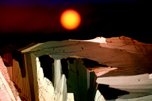
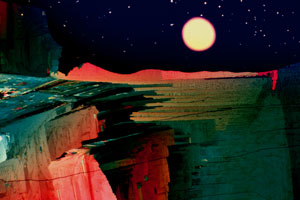
Figure 1.BLUE MOUNTAIN, MONTANA A multiple (4) exposure of ascorbic acid (the plants in the foreground), the biological buffer HEPES (the mountain), stretched polyethylene (the sky), and the field diaphragm image defocused (the sun). |
At the Florida State University, our interests in photomicrography have led us to explore the possibilities of multiple exposure color photomicrography using crystals grown from a wide variety of chemicals, biochemicals, polymers, thin-films, integrated circuits, and biological macromolecules. The basic construction of these photomicrographs involves the classical microscopy techniques of brightfield and cross-polarized illumination, in addition to more modern techniques such as differential interference contrast (Nomarski illumination) and Rheinberg illumination assisted by basic visible light color-filtering processes.
PHOTOMICROGRAPHY AS ART
Using multiple-exposure photomicrography, I have constructed a series of unusual micrographs which I have termed microscapes. Microscapes consist of multiple exposures (usually from 2 to 9) on a single frame of 35 mm transparency film. These photomicrographs are intended to resemble novel alien-like landscapes (see Figures 1-8) and have the highest contrast and color saturation currently available with commercial photographic materials and the E6 process.
|
Figure 2. NEBRASKA A multiple (4) exposure of ascorbic acid (the wheat in the foreground), stretched polyethylene (the morning sky), polybenzyl-l-glutamate spherulites (the stars), and the field diaphragm defocused (the sun). |
The microscape collection of photomicrographs employs both classical and non-classical microscopy techniques. Each micrograph was fabricated using primarily the lower power 4x and 10x objectives where depth-of-field is maximized and the successive exposures remain in focus throughout the field when thin crystalline samples are used. In certain instances, where the illusion of a great distance is intended, a thicker crystal sample or a higher power (20x or 40x) objective with decreased depth-of-field is employed. Under these circumstances, the crystals in the immediate foreground are brought into focus leaving a blurred (out of focus) background to simulate the actual effects generated by depth-of-field limitations with conventional photography equipment.
Selective masking of previously exposed areas is critical, in multiple exposure photography, to avoid undesirable overlap and washing out of successive exposures. Masks can be cut from a portion of black posterboard and are placed over a selected area of the field lens in the base of the microscope, near the field diaphragm.
|
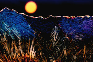
A multiple (4) exposure of the biological buffer HEPES (the foreground and mountains), polybenzyl-l-glutamate spherulites (the stars), stretched polyethylene (the sky), and the field diaphragm image defocused with a yellow filter (the moon). |
Generally, the first step in microscape construction is the exposure of a foreground consisting of a selected recrystallized chemical or biochemical. The foregrounds are imaged with polarized light using the lower power objectives and are positioned in the field so that only the bottom 30-50% of the film is exposed. Unexposed color transparency film is essentially black when processed so that the top half of the film which receives no light will remain black (unexposed) due to the presence of the crossed polarizers. After exposure of the foreground, a selective mask is cut that closely follows the contours of the foreground exposure. This mask is placed on the field lens as described above and successive exposures are made without the first exposure receiving any additional light.
The second exposure can be an additional overlap of the first exposure (see Figure 7, for example), a crystalline formation resembling mountains (Figures 1 and 2), a seascape created with filters (Figure 7), or simply a simulated sky (Figures 2, 4, 5 and 6). Mountains can be simulated using a wide variety of recrystallized chemicals, however certain crystallization patterns appear more realistic than others. The mountain effect in Figure 1 comes from crystals made from the organic biological buffer HEPES (Davidson, 1991a,b). Another useful mountain pattern can be obtained by photographing the edges of cholesteric liquid crystalline domains obtained from concentrated solutions of the polysaccharide xanthin in water (Figure 8). Figure 3 is an example in which the foreground and mountains were composed in a single exposure using an unusual crystalline formation of the buffer HEPES.
|
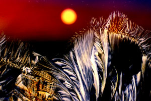
A multiple (4) exposure of recrystallized sulfur (the canyon), polybenzyl-l-glutamate spherulites (the stars), and the field diaphragm defocused (the moon) with a blue filter (the sky). |
I have developed a wide variety of methods for simulating a diversity of sky effects. After carefully trimming a mask to cover all previous exposures, the microscope can be placed in the brightfield mode (by removing the polarizers) and a blue or dark purple filter inserted into the light path (see Figures 5 and 6). Filters which are very light in intensity will work best by allowing the color saturation in the final micrograph to be determined by exposure time. Very short exposures produce an exceptionally deep, dark sky while longer exposures create a much lighter sky. A wide variety of effects can be generated by experimenting with different filters and exposure times. It is very important here that the mask conforms to the topography of all previously exposed regions of the film. On exceedingly irregular boundaries like the mountains in Figures 1, 3, and 8, a very short exposure time will result in a deep blue sky and avoid overlap of blue regions in the mountains.
A spectacular alternative to the blue filter techniques described above involves cutting a 1 x 2 cm section of very thin polyethylene (easily obtained from a sandwich baggy) and stretching it to approximately 1 x 3 cm. The stretched polyethylene is flattened onto a microscope slide and held in place with tape. The act of stretching polyethylene tends to align the chain-like molecules which enhances inherent birefringence in the sample. When viewed through crossed polarizers in the light microscope, the aligned polyethylene molecules combined with the thickness gradient created by stretching causes the sheet of polyethylene to act prism-like. After defocusing the field, sharp features of the polyethylene blend together and the resulting pattern yields a yellow=>red=>blue morning sky effect (Figure 2).
|
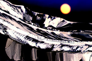
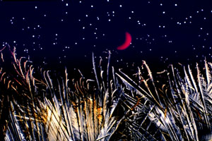
A multiple (4) exposure of ascorbic acid (the wheat in the foreground), polybenzyl-l-glutamate (the stars), the field diaphragm defocused with a mask (the new moon), with a blue sky. |
A third technique which produces an unusual sky effect is the stormy bluish-purple cloudy sky obtained by defocusing a bead of epoxy resin imaged in polarized light with a 530 nm retardation plate positioned between the sample and the analyzer (Figure 6). Light diffracted by the foreground mask accentuates the highlights producing an uncanny storm-like appearance. A blue filter can be clipped from a sheet of dyed acetate and added to the edge of the mask to make the effect more realistic (not illustrated).
After completion of the sky, a sun or moon can be added to the micrograph. This is accomplished by closing the field diaphragm until the desired sun or moon diameter is reached. Next, the image of the shutter leaves is defocused, by lowering the condenser from its "Köhler" position, until they merge to form a smooth circle. After placing the appropriate filter (usually a deep orange, red, or yellow) in the light path, the image of the field diaphragm can be relocated to any position in the field simply by adjusting the centering thumbscrews located on the substage condenser. Placing a mask over the bottom portion of the diaphragm image will create a rising sun effect as illustrated in Figures 6 and 7. Exposure times dictate the color distribution effects obtained with the various filters placed in the light path. With the yellow or orange filters, exceedingly short exposure times produce images with a reddish perimeter and a very color-saturated yellowish interior (Figures 1-3) while longer exposure times tend to wash out the image to yield more of a moon-like effect illustrated in Figure 1. Long exposure times when using a red filter will cause a yellowish center (Figure 6), while shorter times result in a more uniform color effect (not illustrated).
![]()
|
A multiple exposure (4) of ascorbic acid (the desert foreground), stretched polyethylene (the morning sky), polybenzyl-l-glutamate (the stars), and the field diaphragm defocused (the rising sun). |
To simulate the reflection of the sun or moon on water, two methods can be used. After the field diaphragm image is correctly positioned in the field, a diffraction grating can be inserted into the light path to spread the image and shift the colors to longer (redder) wavelengths as illustrated in Figure 7. I typically use Polachrome HC instant 35 mm transparency film exposed to intense daylight because the manufacturing process for this film results in a series of very finely spaced lines which serve as an excellent diffraction grating when the film is greatly overexposed. Alternatively, a fine-tooth comb inserted into the light path will produce the effect shown in Figure 7.
The new moon concept depicted in Figure 5 is produced simply by inserting the tip of a ball-point pen into the light path after the field diaphragm is defocused and correctly positioned. A yellow filter is used here instead of orange to produce a more realistic appearance.
|
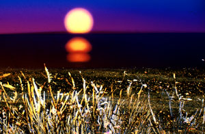
A multiple (6) exposure of ascorbic acid (two exposures: the sandy beach and the sea oats in the foreground), stretched polyethylene with a blue filter (the morning sky and ocean), Cibachrome bleach crystallites defocused (the clouds), and the field diaphragm image defocused with a yellow filter (two exposures: the rising sun and it's reflection). |
Generation of stars and/or clouds is the final exposure in the sequence. After the field diaphragm exposure has been completed, a sealed microscope slide containing a solution of small liquid crystalline spherulites (spherical structures approximately 2µ in diameter) of the rod-like a-helical polypeptide polybenzyl-L-glutamate is placed on the microscope stage. At relatively low magnifications (using the 10x objective), the spherulites appear as point-sources of light (Figures 2-5). When the desired field of spherulites is imaged, the field diaphragm can be closed again until the area visible in the field is equal to the diameter of the sun or moon produced by the previous exposure. By carefully manipulating the position of the polybenzyl-L-glutamate spherulite microslide, an area on the slide can be located which is devoid of spherulites and this area is placed directly over the previously exposed field diaphragm image area. This prevents stars from being imaged in the center of the moon or sun. Clouds are produced by defocusing colorless birefringent crystallites. Several chemicals produce the needed crystallites including Cibachrome bleach crystals (Figures 7 and 8), most black and white paper developers, and a variety of laboratory reagents and sugars. The crystals are usually imaged in the top-most portion of the viewfield and exposed for long times to wash out all color.
|
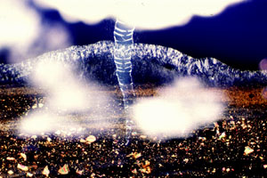
A multiple (9) exposure of ascorbic acid (Vitamin C-the desert foreground), cholesteric liquid crystalline DNA (the tornado), Xanthan-gum (the mountains), and Cibachrome bleach crystals (the clouds and dust) with a blue sky. |
Perhaps the most difficult shot is the beach scene (Figure 7) where the morning sky and ocean are created in a single exposure. This is accomplished by placing a blue filter over the foreground mask at the field lens and defocusing the microscope until a sharp boundary occurs at the junction of the blue filter and the stretched polyethylene image. Correct positioning of the field diaphragm image to create a rising sun effect can be especially difficult when attempting to construct beach scenes.
CONCLUSIONS
It is not necessary to follow the diagrammed outline exactly in order to obtain nicely formed microscapes. The most important aspect is correct positioning of each successive exposure regardless of the order in which the exposures are recorded. This aspect of the multiple exposure photomicrography can be assisted by the use of a graded photography reticle in the ocular of the microscope. By allowing a small degree of overlap between the exposures (approximately 5%), a blending of the features occurs which lends a touch of reality to the microscapes. The number of crystallizable chemicals which will prove beneficial in the construction of microscapes is probably limitless and, by trial and error, a very broad spectrum of different microscapes is no doubt possible. A nice "fringe benefit" of this type of work is that the interested photomicrographer can fine-tune his or her photomicrographic skills. With the proper equipment on hand for the assembly of microscapes, the interested microscopist is limited only by the boundaries of the imagination.
ACKNOWLEDGEMENTS
I would like to thank Jit Gurung for assistance with color photography techniques and the FSU Center for Materials Research and Technology (MARTECH) for continued support. Stephen Page and Lynda Ruf from the Nikon Instrument Group provided all photomicrography equipment used in this research.
REFERENCES
Davidson, M. W., (1991a) "Fabrication of Unusual Art Forms with Multiple Exposure Color Photomicrography" the MICROSCOPE 38: 357-369.
Davidson, M. W., (1991b) "Fascinating Photography with a Simple Light Microscope" PETERSON'S PHOTOGRAPHIC April pages 92-97.
About the Author: Michael W. Davidson is a research scientist with the National High Magnetic Field Laboratory and the Supercomputer Computations Research Institute at the Florida State University in Tallahassee. His main research interests are liquid crystalline biological systems and the packaging of DNA in virus heads. Davidson has published over 75 scientific and popular magazine articles and has had his photomicrographs published on the covers of over a 100 journals and magazines.