Fluorescence Digital Image Gallery
Horse Dermal Fibroblast Cells (NBL-6)
|
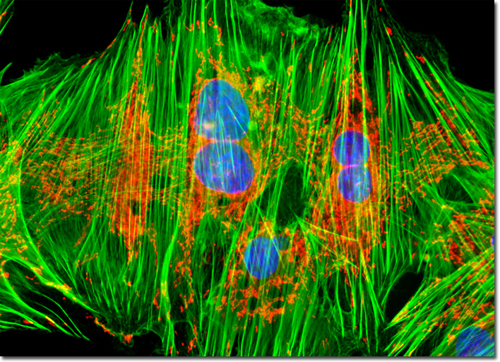
The Alexa Fluor series of dyes are commercially available as reactive intermediates in the form of maleimides, succinimidyl esters, and hydrazides, as well as prepared cytoskeletal probes and conjugates, providing a broad array of tools for fluorescence and immunofluorescence applications. For researchers needing to visualize the filamentous actin elements of the cytoskeleton, Alexa Fluor dyes conjugated to a number of fluorescent and biotinylated derivatives of phalloidin and phallacidin have been designed. These phallotoxins are derived from the toxic death cap mushroom (Amanita phalloides) and competitively bind to the same sites on F-actin. The Alexa Fluor dye-labeled phallotoxins are equally selective for both large and small filaments, and do not bind to the monomeric form of actin (G-actin), as do some antibodies against actin. Moreover, unlike antibodies, the binding characteristics of the phallotoxins do not significantly alter with actin from different species. The culture of NBL-6 horse fibroblasts illustrated above was stained with MitoTracker Red CMXRos and Alexa Fluor 488 (green emission) conjugated to phalloidin in order to target mitochondria and the cytoskeletal F-actin network, respectively. In addition, nuclear DNA was labeled with Hoechst 33342 (blue emission). Images were recorded in grayscale with a QImaging Retiga Fast-EXi camera system coupled to an Olympus BX-51 microscope equipped with bandpass emission fluorescence filter optical blocks provided by Omega Optical. During the processing stage, individual image channels were pseudocolored with RGB values corresponding to each of the fluorophore emission spectral profiles. |
© 1995-2025 by Michael W. Davidson and The Florida State University. All Rights Reserved. No images, graphics, software, scripts, or applets may be reproduced or used in any manner without permission from the copyright holders. Use of this website means you agree to all of the Legal Terms and Conditions set forth by the owners.
This website is maintained by our
|