
Fluorescence Digital Image Gallery
Horse Dermal Fibroblast Cells (NBL-6)
The NBL-6 cell line, also known as E. Derm, was derived from the dermis of a 4-year-old female horse (Equus caballus) of the quarterhorse strain. The cells, which appear to senesce after approximately 40 passages, are susceptible to herpes simplex, reovirus 3, vesicular stomatitis (Ogden strain), and vaccinia, but resist adenovirus 5, coxsackievirus A9 and B5, and poliovirus 2.
NBL-6 cells, which are negative for reverse transcriptase and exhibit typical fibroblast morphology, have been utilized for a variety of studies, as well as for virus propagation for equine vaccines. The cell line has been particularly important in research focusing upon equine viral arteritis (EVA), a contagious viral disease that affects horse populations worldwide and appears to be increasing in incidence.
Derived from the mesoderm, the dermis is a dense layer of connective tissue that underlies the epidermis. The interface between these two strata is comprised of dermal ridges interlocked with epidermal ridges, creating an irregular pattern that can be observed along the surface of the skin, especially at the palms of the hands and soles of the feet. The dermis, which is predominantly comprised of fibroblasts but also contains mast cells, macrophages, and other cell types associated with connective tissue, is typically considered to be subdivided into two zones: a papillary layer and a reticular layer. Composed of more loosely arranged collagen and elastic fibers than the underlying dense reticular zone, the papillary layer exhibits a number of capillary loops that function in the regulation of body temperature and the nourishment of epidermal cells. Within the thick network of collagen fibers characteristic of the reticular layer, however, a number of other important structures reside, including sweat glands, sebaceous glands, hair follicles, and mechanoreceptors.
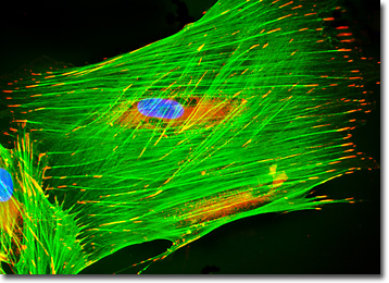
The culture of horse dermal fibroblast cells presented in the digital image above was immunofluorescently labeled with anti-vinculin mouse monoclonal primary antibodies followed by goat anti-mouse Fab fragments conjugated to Alexa Fluor 568 (yielding red emission). In addition, the specimen was simultaneously stained for DNA with Hoechst 33342 (blue emission), and for the cytoskeletal filamentous actin network with Alexa Fluor 488 (green emission) conjugated to phalloidin. Images were recorded in grayscale with a QImaging Retiga Fast-EXi camera system coupled to an Olympus BX-51 microscope equipped with bandpass emission fluorescence filter optical blocks provided by Omega Optical. During the processing stage, individual image channels were pseudocolored with RGB values corresponding to each of the fluorophore emission spectral profiles.
Additional Fluorescence Images of Horse Dermal Fibroblast (NBL-6) Cells
Mitochondrial Distribution in Horse (NBL-6) Fibroblasts - The mitochondrial network was targeted in a culture of NBL-6 horse fibroblasts with the X-rosamine derivative, MitoTracker Red CMXRos. The culture was simultaneously labeled for the cytoskeletal filamentous actin network and nuclear DNA with Alexa Fluor 488 conjugated to phalloidin and Hoechst 33258, respectively.
Targeting Intermediate Filaments and the Golgi Network in NBL-6 Cells with Immunofluorescence - NBL-6 dermal fibroblast cells were fixed with paraformaldehyde, permeabilized, and treated with a mixture of rabbit (anti-giantin) and mouse (anti-vimentin; pan) primary antibodies, followed by secondary antibodies conjugated to Oregon Green 488 and Texas Red, respectively. Cell nuclei were counterstained with the DNA-selective bisbenzimide dye, Hoechst 33342.
Horse Dermal (NBL-6) Cells with MitoTracker Red CMXRos, Alexa Fluor 488, and Hoechst 33342 - A culture of NBL-6 horse fibroblasts was stained with MitoTracker Red CMXRos and Alexa Fluor 488 (green emission) conjugated to phalloidin in order to target mitochondria and the cytoskeletal F-actin network, respectively. In addition, nuclear DNA was labeled with Hoechst 33342 (blue emission).
Labeling the Endoplasmic Reticulum in Fibroblast Cultures with Immunofluorescence - Calreticulin is a calcium-binding protein resident in the membrane of the endoplasmic reticulum. The adherent culture of horse fibroblasts featured in this section was immunofluorescently labeled with anti-calreticulin primary rabbit monoclonal antibodies followed by goat anti-rabbit secondary antibodies conjugated to Cy3. The cells were also stained with Alexa Fluor 488 conjugated to phalloidin and Hoechst 33342, which bind respectively with filamentous actin and DNA in the cell nucleus.
Microtubular and Golgi Networks in Dermal Fibroblasts - In order to visualize the relationship between the Golgi complex and the intracellular microtubular network, a culture of horse dermal fibroblast (NBL-6) cells was immunofluorescently labeled with anti-tubulin (pan) mouse monoclonal primary antibodies followed by goat anti-mouse Fab fragments conjugated to Alexa Fluor 488. Golgi bodies were simultaneously targeted with rabbit anti-giantin primary antibodies, followed by goat anti-rabbit secondaries conjugated to Alexa Fluor 568. Nuclei were counterstained with Hoechst 33342.
BACK TO THE CULTURED CELLS FLUORESCENCE GALLERY
BACK TO THE FLUORESCENCE GALLERY