Fluorescence Digital Image Gallery
Madin-Darby Ovine Kidney Epithelial Cells (MDOK)
|
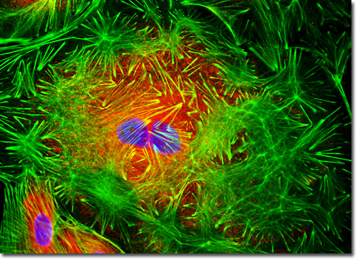
The endoplasmic reticulum (ER) is an extensive net-like membrane complex present in eukaryotic cells consisting of bifurcating tubules and the sac-like structures known as cisternae. In many animal cells, the organelle accounts for more than 50 percent of the total amount of membranous material present in the cytoplasm. The variously shaped components of the organelle are interconnected, and together separate the highly convoluted internal region of the endoplasmic reticulum, often referred to as the lumen or cisternal space, from the surrounding cytosol. The endoplasmic reticulum membrane is also contiguous with the envelope that encases the nucleus, thus, the region between the two membranes that comprise the nuclear envelope is continuous with ER lumen. In addition to significantly compartmentalizing the cell and functioning as a selective barrier, the membrane of the endoplasmic reticulum carries out a number of other important functions, such as the synthesis of lipids and proteins as well as the detoxification of poisonous substances. The fixed and permeabilized adherent culture of MDOK cells illustrated above was treated with a cocktail of concanavalin A conjugated to Texas Red and Alexa Fluor 488 conjugated to phalloidin to label the endoplasmic reticulum and filamentous actin networks, respectively. The nuclei were subsequently counterstained with Hoechst 33258. Images were recorded in grayscale with a QImaging Retiga Fast-EXi camera system coupled to an Olympus BX-51 microscope equipped with bandpass emission fluorescence filter optical blocks provided by Omega Optical. During the processing stage, individual image channels were pseudocolored with RGB values corresponding to each of the fluorophore emission spectral profiles. |
© 1995-2025 by Michael W. Davidson and The Florida State University. All Rights Reserved. No images, graphics, software, scripts, or applets may be reproduced or used in any manner without permission from the copyright holders. Use of this website means you agree to all of the Legal Terms and Conditions set forth by the owners.
This website is maintained by our
|