Fluorescence Digital Image Gallery
Madin-Darby Canine Kidney Epithelial Cells (MDCK)
|
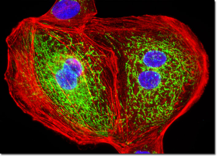
In mammalian mitochondria, the process of oxidative phosphorylation (often abbreviated with the acronym OxPhos) is catalyzed by five large membrane-bound protein complexes that are the targets of a wide variety of monoclonal antibodies. All of these complexes are composed of multiple protein subunits that are encoded by either the mitochondrial DNA or the nuclear DNA. As an example, mammalian cytochrome oxidase consists of 13 subunits, with three of the sequences deriving from the mitochondria and 10 from the nucleus. Mitochondrial oxidative phosphorylation complex V is also a multi-subunit combination that contains an ATP synthase subunit coupled to a proton channel. An inhibitor protein that acts on the ATPase moiety is not considered a member of complex V, but does associate with this OxPhos subsystem. Antibodies targeting the ATPase inhibitor protein are good candidates for immunofluorescence localization of the mitochondria in fluorescence microscopy. The adherent monolayer culture of Madin-Darby canine kidney cells illustrated above was immunofluorescently labeled with primary mouse anti-oxphos complex V inhibitor protein antibodies, followed by goat anti-mouse Fab fragments conjugated to fluorescein. The culture was subsequently stained with Alexa Fluor 568 conjugated to phalloidin to reveal details of the filamentous actin network, and DAPI for DNA in the nucleus. Images were recorded in grayscale with a QImaging Retiga Fast-EXi camera system coupled to an Olympus BX-51 microscope equipped with bandpass emission fluorescence filter optical blocks provided by Omega Optical. During the processing stage, individual image channels were pseudocolored with RGB values corresponding to each of the fluorophore emission spectral profiles. |
© 1995-2025 by Michael W. Davidson and The Florida State University. All Rights Reserved. No images, graphics, software, scripts, or applets may be reproduced or used in any manner without permission from the copyright holders. Use of this website means you agree to all of the Legal Terms and Conditions set forth by the owners.
This website is maintained by our
|