Fluorescence Digital Image Gallery
Normal African Green Monkey Kidney Fibroblast Cells (CV-1)
|
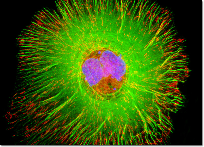
In 1967, only three years after the CV-1 cell line was first established, an acute, highly fatal hemorrhagic fever caused by an agent that would come to be known as the Marburg virus broke out in Germany among scientists and technicians working with the tissues and blood of African green monkeys that had been shipped from Uganda. In this instance, more than 30 individuals were affected, many of which did not survive. Since that time, several other instances of the Marburg hemorrhagic fever have been documented, but these have only involved small numbers of people. In a typical outbreak, the first to become infected is someone who has spent a significant amount of time traveling in Africa, where the virus is indigenous. Subsequently, health care professionals, family members, or others that come into contact with the infected individual may become sick as well. The exact mode of initial transmission of the virus from an animal host is not known, but secondary transmission may be prevented through barrier nursing techniques that enable caregivers to avoid direct contact with the patient. The isolated fibroblast that appears in the digital image above was resident in a normal (non-transformed) African green monkey kidney cell culture labeled with MitoTracker Red CMXRos, Alexa Fluor 488 conjugated to phalloidin, and DAPI, targeting the mitochondrial network, F-actin, and nuclear DNA, respectively. Note the striking geometry of actin filaments, which demonstrate stress fibers, lamellipodia, filopodia, microspikes, and dorsal arcs. Images were recorded in grayscale with a QImaging Retiga Fast-EXi camera system coupled to an Olympus BX-51 microscope equipped with bandpass emission fluorescence filter optical blocks provided by Omega Optical. During the processing stage, individual image channels were pseudocolored with RGB values corresponding to each of the fluorophore emission spectral profiles. |
© 1995-2025 by Michael W. Davidson and The Florida State University. All Rights Reserved. No images, graphics, software, scripts, or applets may be reproduced or used in any manner without permission from the copyright holders. Use of this website means you agree to all of the Legal Terms and Conditions set forth by the owners.
This website is maintained by our
|