Fluorescence Digital Image Gallery
Normal African Green Monkey Kidney Fibroblast Cells (CV-1)
|
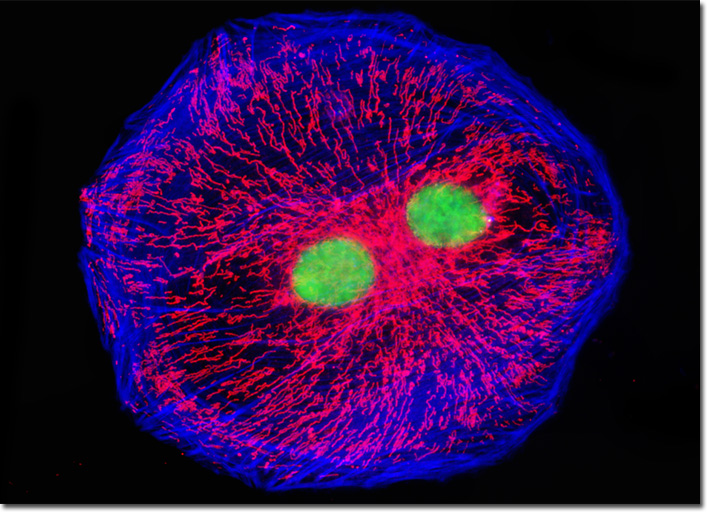
According to the traditional view of fibroblasts, all of the cells are similar in morphology and function. Modern studies, however, have provided ample evidence demonstrating that this is not a true characterization of fibroblasts. Instead, the prevailing view of the cells in recent years has been that some subsets of fibroblasts are able to give rise to other types of cells, such as bone cells, fat cells, and smooth muscle cells. Due to this change in perception, the scientific community is also beginning to discover that fibroblasts are more heavily involved in certain diseases and medical conditions than previously supposed. For example, scarring occasionally appears around internal organs without any evidence of tissue damage, and kidney fibroblasts, such as those that comprise the CV-1 cell line, may be involved in the unusual development. Over time, this fresh attention to fibroblasts may eventually lead to the development of new kinds of medications that help impede the excessive buildup of scar tissue, fat cells, or other kinds of matter that may be produced by the cells. The isolated cell that appears in the digital image above was a resident in an adherent monolayer culture of CV-1 cells labeled with SYTOX Green to stain chromatin in the nuclei. In addition, the culture was treated with Alexa Fluor 350 conjugated to phalloidin and MitoTracker Red CMXRos, to target filamentous actin and mitochondrial network, respectively. Images were recorded in grayscale with a QImaging Retiga Fast-EXi camera system coupled to an Olympus BX-51 microscope equipped with bandpass emission fluorescence filter optical blocks provided by Omega Optical. During the processing stage, individual image channels were pseudocolored with RGB values corresponding to each of the fluorophore emission spectral profiles. |
© 1995-2025 by Michael W. Davidson and The Florida State University. All Rights Reserved. No images, graphics, software, scripts, or applets may be reproduced or used in any manner without permission from the copyright holders. Use of this website means you agree to all of the Legal Terms and Conditions set forth by the owners.
This website is maintained by our
|