Fluorescence Digital Image Gallery
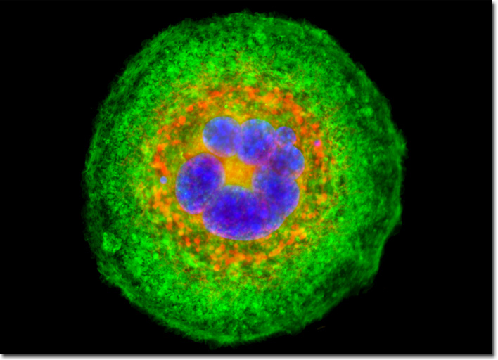
Transformed Mouse Cerebellum Microglial Cells (C8-B4)
|
Mitochondrial probes are among the most useful fluorophores for investigating cellular respiration and are often employed along with other dyes in multiple labeling investigations. Some of the traditional dyes utilized for such purposes include Rhodamine 123 and tetramethylrosamine. However, these probes are rapidly lost when cells are fixed and, therefore, have largely been supplanted by newer, more specific, fluorophores, including those of the MitoTracker series. MitoTracker probes, which are available in a variety of excitation and emission spectral profiles, contain a mildly thiol-reactive chloromethyl moiety. Apparently, the presence of the chloromethyl group is involved in maintaining the dye's association with cellular mitochondria subsequent to fixation. The C8-B4 cell that appears in the digital image above was resident in a culture of mouse microglial cells labeled with MitoTracker Red CMXRos, targeting the intracellular mitochondrial network. In addition, the cells were labeled for filamentous actin and DNA in the nucleus with Alexa Fluor 488 conjugated to phalloidin and Hoechst 33258, respectively. Images were recorded in grayscale with a QImaging Retiga Fast-EXi camera system coupled to an Olympus BX-51 microscope equipped with bandpass emission fluorescence filter optical blocks provided by Omega Optical. During the processing stage, individual image channels were pseudocolored with RGB values corresponding to each of the fluorophore emission spectral profiles. |
© 1995-2025 by Michael W. Davidson and The Florida State University. All Rights Reserved. No images, graphics, software, scripts, or applets may be reproduced or used in any manner without permission from the copyright holders. Use of this website means you agree to all of the Legal Terms and Conditions set forth by the owners.
This website is maintained by our
|