
Fluorescence Digital Image Gallery
Transformed Mouse Cerebellum Microglial Cells (C8-B4 Line)
Microglia are specialized macrophages that are very important for their role in protecting the central nervous system. C8-B4 is a spontaneously transformed microglial clone of a cell line originally derived from the cerebellum of an 8-day-old mouse (Mus musculus) in 1984. This initial organ culture was used to establish several distinct astroglial cell lines.
The C8-B4 clone was created in 1996, and the neuronal cells grow adherently in culture. Classical microglial markers, including MAC1, F4/80, and 2-4G2, are expressed by the C8-B4 clone, which appears to be derived from a microglial precursor since it reportedly does not express differentiation antigens present during the early stage of the monocytic lineage. C8-B4 cells produce and release large amounts of glutamate, a substance that typically functions as a neurotransmitter.
C8-B4 cells also notably synthesize CD4, a molecule of intense scientific interest due to its role in the proliferation of the human immunodeficiency virus (HIV). As a retrovirus, HIV is forced to rely upon the host cell to complete viral replication. More specifically, a protein called gp120 embedded in the envelope surrounding the genetic information of HIV binds to CD4 located on the surface of a helper T cell. When this occurs, a series of reactions follows that enable the genetic information of HIV to enter into the cell. One of the most important of these reactions appears to be the exposure of a region of gp120 due to the CD4-virus bond that elicits a conformational change that exposes part of gp41, a viral envelope protein, which introduces itself into the host cell membrane, essentially bridging the gap between viral envelope and the membrane. Following additional changes in the gp41 protein, fusion between the envelope and membrane typically occurs and the genetic information of HIV enters the host cell.
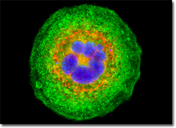
The C8-B4 cell that appears in the digital image above was resident in a culture of mouse microglial cells labeled with MitoTracker Red CMXRos, targeting the intracellular mitochondrial network. In addition, the cells were labeled for filamentous actin and DNA in the nucleus with Alexa Fluor 488 conjugated to phalloidin and Hoechst 33258, respectively. Images were recorded in grayscale with a QImaging Retiga Fast-EXi camera system coupled to an Olympus BX-51 microscope equipped with bandpass emission fluorescence filter optical blocks provided by Omega Optical. During the processing stage, individual image channels were pseudocolored with RGB values corresponding to each of the fluorophore emission spectral profiles.
BACK TO THE CULTURED CELLS FLUORESCENCE GALLERY
BACK TO THE FLUORESCENCE GALLERY