Fluorescence Digital Image Gallery
Human Lung Carcinoma Cells (A-549)
|
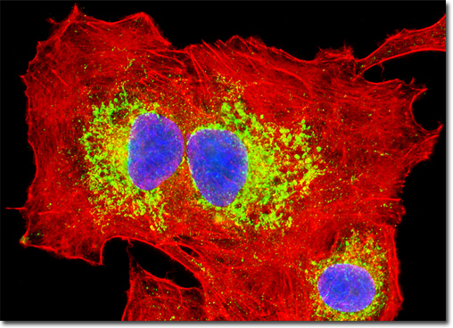
The A-549 cell line was originally cultivated in 1972 by D. J. Giard, along with several collaborators, from the human lung carcinoma of a 58-year-old Caucasian male. The line is commonly used to investigate a wide range of respiratory ailments, such as viral infections capable of inducing asthma, tissue damage linked to asbestos exposure, and smoking-related emphysema. Adherent and epithelial, A-549 cells are positive for keratin by immunoperoxidase staining, but are negative for reverse transcriptase, indicating the lack of integral retrovirus genomes. Studies by a team led by M. Lieber have revealed that A-549 cells are able to synthesize lecithin with a high percentage of desaturated fatty acids utilizing the cytidine diphosphocholine pathway. The culture of human carcinoma (A-549) cells presented in the digital image above was immunofluorescently labeled with primary anti-oxphos complex V inhibitor protein monoclonal antibodies (mouse) followed by goat anti-mouse Fab fragments conjugated to Alexa Fluor 488. The cells were also stained for F-actin with Alexa Fluor 568 conjugated to phalloidin, and for DNA in the nucleus with DAPI. Images were recorded in grayscale with a QImaging Retiga Fast-EXi camera system coupled to an Olympus BX-51 microscope equipped with bandpass emission fluorescence filter optical blocks provided by Omega Optical. During the processing stage, individual image channels were pseudocolored with RGB values corresponding to each of the fluorophore emission spectral profiles. |
© 1995-2025 by Michael W. Davidson and The Florida State University. All Rights Reserved. No images, graphics, software, scripts, or applets may be reproduced or used in any manner without permission from the copyright holders. Use of this website means you agree to all of the Legal Terms and Conditions set forth by the owners.
This website is maintained by our
|