
Fluorescence Digital Image Gallery
Human Lung Carcinoma Cells (A-549 Line)
The A-549 cell line was originally cultivated in 1972 by D. J. Giard, along with several collaborators, from the human lung carcinoma of a 58-year-old Caucasian male. The line is commonly used to investigate a wide range of respiratory ailments, such as viral infections capable of inducing asthma, tissue damage linked to asbestos exposure, and smoking-related emphysema.
Adherent and epithelial, A-549 cells are positive for keratin by immunoperoxidase staining, but are negative for reverse transcriptase, indicating the lack of integral retrovirus genomes. Studies by a team led by M. Lieber have revealed that A-549 cells are able to synthesize lecithin with a high percentage of desaturated fatty acids utilizing the cytidine diphosphocholine pathway.
A carcinoma, such as the one that served as the original source of the A-549 cell line, is a solid, cancerous growth that begins its development in the epithelial tissue that forms human skin and comprises the linings of most organs and glands. The cells of a carcinoma may spread, however, to nearby tissues that are otherwise healthy and can also generate secondary tumors known as metastases in parts of the body that are distant from the initial growth. Some of the most common areas of carcinoma development include the skin, lungs, uterus, stomach, ovaries, and prostate, though the incidence of growths in these and other areas vary significantly by country. Lung carcinoma, which was deemed relatively rare in the early twentieth century, is currently diagnosed more than any other form of major cancer worldwide. It is also the most common cause of cancer fatalities in both men and women. In countries where cigarette smoking has been prevalent for many years, as many as 90 percent of patients diagnosed with lung cancer are, or have been, smokers.
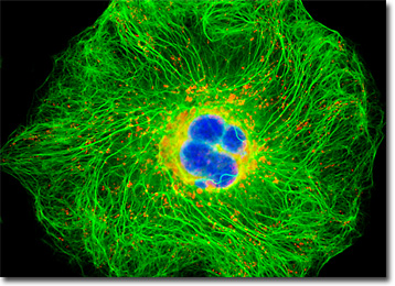
The single human carcinoma cell illustrated above was resident in a culture that was immunofluorescently labeled with primary anti-cytokeratin (pan) mouse monoclonal antibodies followed by goat anti-mouse Fab fragments conjugated to the cyanine probe, Cy2. In addition, the specimen was treated with MitoTracker Red CMXRos and Hoechst 33258 to stain the mitochondrial network and nuclei, respectively. Images were recorded in grayscale with a QImaging Retiga Fast-EXi camera system coupled to an Olympus BX-51 microscope equipped with bandpass emission fluorescence filter optical blocks provided by Omega Optical. During the processing stage, individual image channels were pseudocolored with RGB values corresponding to each of the fluorophore emission spectral profiles.
Additional Fluorescence Images of Human Lung Carcinoma (A-549) Cells
A-549 Cells with Alexa Fluor 568, Alexa Fluor 488, and DAPI - An adherent culture of A-549 cells was stained for F-actin with Alexa Fluor 568 conjugated to phalloidin, and for DNA with 4',6-diamidino-2-phenylindole (DAPI). The cells were additionally labeled for lipid vesicles with Alexa Fluor 488 conjugated to lectin PNA, a peanut-derived protein that binds to specific carbohydrate groups on proteins or cell membranes.
Cytokeratin in Human Lung Carcinoma Cells - A human lung carcinoma (A-549) cell culture was immunofluorescently labeled with primary anti-cytokeratin (an intermediate filament protein) mouse monoclonal antibodies followed by goat anti-mouse Fab fragments conjugated to Marina Blue. The culture was also stained with MitoTracker Red CMXRos and SYTOX Green to probe the mitochondrial network and nuclei, respectively.
A-549 Cells with Alexa Fluor 488, Alexa Fluor 350, and Propidium Iodide - The adherent culture of human carcinoma (A-549) cells illustrated in this section was stained with Alexa Fluor 350 conjugated to wheat germ agglutinin for the Golgi apparatus, as well as Alexa Fluor 488 conjugated to phalloidin for cytoskeletal actin. Nuclei were labeled with the intercalating dye, propidium iodide.
Enhanced Yellow Protein (EYFP) Subcellular Localization in A-549 Human Lung Carcinoma Cells - A log phase culture of A-549 cells was transfected with an pEYFP-Mitochondria plasmid subcellular localization vector, which contains the mitochondrial targeting sequence from subunit VIII of human cytochrome C oxidase. The enhanced yellow fluorescent protein gene employed with this culture features several important amino acid substitutions that shift the emission maximum of green fluorescent protein (GFP) by approximately 18 nanometers, from 509 to 527 nanometers. The cells were additionally labeled with Alexa Fluor 568 conjugated to phalloidin and DAPI, targeting the filamentous actin network and nuclei, respectively.
A-549 Cells with MitoTracker Red CMXRos, Marina Blue, and SYTOX Green - A-549 cells were immunofluorescently labeled with primary anti-cytokeratin (an intermediate filament protein) mouse monoclonal antibodies followed by goat anti-mouse Fab fragments conjugated to Marina Blue. MitoTracker Red CMXRos (mitochondria) and SYTOX Green (nuclei) were also used to counterstain the carcinoma cell culture.
Human Lung Carcinoma Cells with Cy2, MitoTracker Red CMXRos, and Hoechst 33258 - The human lung carcinoma cell presented in this section was resident in a culture immunofluorescently labeled with primary anti-cytokeratin (pan) mouse monoclonal antibodies followed by goat anti-mouse Fab fragments conjugated to Cy2. In addition, the specimen was stained with MitoTracker Red CMXRos and Hoechst 33258 to label the mitochondrial network and nuclei, respectively.
A-549 Cells with Alexa Fluor 568, Alexa Fluor 488, and DAPI - A culture of human lung carcinoma (A-549) cells was immunofluorescently labeled with primary anti-oxphos complex V inhibitor protein monoclonal antibodies (mouse) followed by goat anti-mouse Fab fragments conjugated to Alexa Fluor 488. The cells were also stained for F-actin with Alexa Fluor 568 conjugated to phalloidin, and for DNA in the nucleus with DAPI.
DsRed Fluorescent Protein in Transfected Human Lung Carcinoma Cell Cultures - After transiently transfecting a log phase culture of A-549 cells (DsRed2-Mitochondria; 10-percent transfection efficiency), the culture was washed, fixed, permeabilized, and blocked with bovine serum albumen. The cells were subsequently labeled with Alexa Fluor 488 conjugated to phalloidin and counterstained with DAPI. Note that the two central cells in the image are the only individuals expressing the chimeric protein.
Histone and Peroxisome Distribution in A-549 Cell Cultures - In a double immunofluorescence experiment, an adherent culture of human lung carcinoma epithelial cells was fixed, permeabilized, blocked with 10 percent normal goat serum, and treated with a cocktail of mouse anti-histones (pan) and rabbit anti-PMP 70 (peroxisomal membrane protein) primary antibodies, followed by goat anti-mouse and anti-rabbit secondary antibodies (IgG) conjugated to Texas Red and Alexa Fluor 488, respectively. The filamentous actin network was counterstained with Alexa Fluor 350 conjugated to phalloidin.
Cytokeratin Intermediate Filaments and the Golgi Network in Human Lung Carcinoma Cells - In order to visualize the relationship between the Golgi complex and the cytokeratin intermediate filament network, a culture of human lung carcinoma (A-549) cells was immunofluorescently labeled primary anti-cytokeratin (pan) mouse monoclonal antibodies followed by goat anti-mouse Fab fragments conjugated Texas Red. Golgi bodies were simultaneously targeted with rabbit anti-giantin primary antibodies, followed by goat anti-rabbit secondaries conjugated to Oregon Green 488. Nuclei were counterstained with Hoechst 33342.
Focal Adhesion Sites in Human Lung Carcinoma Epithelial Cells - A culture of A-549 cells was immunofluorescently labeled with primary anti-vinculin mouse monoclonal antibodies followed by goat anti-mouse secondary antibodies (IgG) conjugated to Alexa Fluor 568 (red fluorescence emission). Note the prominent staining of the cellular attachment network in the central portion and periphery of these cells. In addition, the specimen was simultaneously stained for DNA with the ultraviolet-absorbing probe Hoechst 33258, and for the cytoskeletal filamentous actin network with Alexa Fluor 488 conjugated to phalloidin.
Labeling the Mitochondrial Network in A-549 Cells with Enhanced Yellow Fluorescent Protein - This section features a culture of human lung carcinoma cells that was transfected with a pEYFP-Mitochondria plasmid subcellular localization vector, which contains the mitochondrial targeting sequence from subunit VIII of human cytochrome C oxidase. The enhanced yellow fluorescent protein gene used with this culture contains several important amino acid substitutions that shift the emission maximum of green fluorescent protein (GFP) by approximately 18 nanometers, from 509 to 527 nanometers. The cells were additionally labeled with the nucleic acid stain SYTOX Orange and Alexa Fluor 350 conjugated to phalloidin, targeting DNA and filamentous actin, respectively.
BACK TO THE CULTURED CELLS FLUORESCENCE GALLERY
BACK TO THE FLUORESCENCE GALLERY