Fluorescence Digital Image Gallery
Human Lung Carcinoma Cells (A-549)
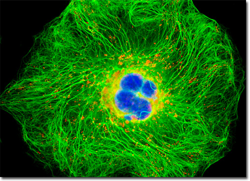
|
The tumors that may plague the lungs are typically classified into two basic types: small-cell carcinomas and non-small-cell carcinomas. The small-cell type, which accounts for approximately a quarter of diagnosed cases, is the more aggressive of the two and rarely occurs in people who have never been smokers. The cells of a small-cell carcinoma, as is suggested by the tumor name, are diminutive in size, as well as round, oval, or oat-like in shape. Non-small-cell carcinomas, which occur much more frequently than the small-cell variety, are generally subdivided into three primary kinds: squamous cell carcinomas, adenocarcinomas, and large-cell carcinomas. The most common of these in the United States is adenocarcinoma, the cells of which are cube- or column-shaped. Squamous cell carcinoma, which is only slightly less common than adenocarcinoma, features cells that are flat and scale-like. Large-cell carcinomas do not have a characteristic cell shape, however, since any tumor of the lung that does not fit into any of the other classifications are generally placed in this group. The single human carcinoma cell illustrated above was resident in a culture that was immunofluorescently labeled with primary anti-cytokeratin (pan) mouse monoclonal antibodies followed by goat anti-mouse Fab fragments conjugated to the cyanine probe, Cy2. In addition, the specimen was treated with MitoTracker Red CMXRos and Hoechst 33258 to stain the mitochondrial network and nuclei, respectively. Images were recorded in grayscale with a QImaging Retiga Fast-EXi camera system coupled to an Olympus BX-51 microscope equipped with bandpass emission fluorescence filter optical blocks provided by Omega Optical. During the processing stage, individual image channels were pseudocolored with RGB values corresponding to each of the fluorophore emission spectral profiles. |
© 1995-2025 by Michael W. Davidson and The Florida State University. All Rights Reserved. No images, graphics, software, scripts, or applets may be reproduced or used in any manner without permission from the copyright holders. Use of this website means you agree to all of the Legal Terms and Conditions set forth by the owners.
This website is maintained by our
|