Fluorescence Digital Image Gallery
Embryonic Swiss Mouse Fibroblast Cells (3T3)
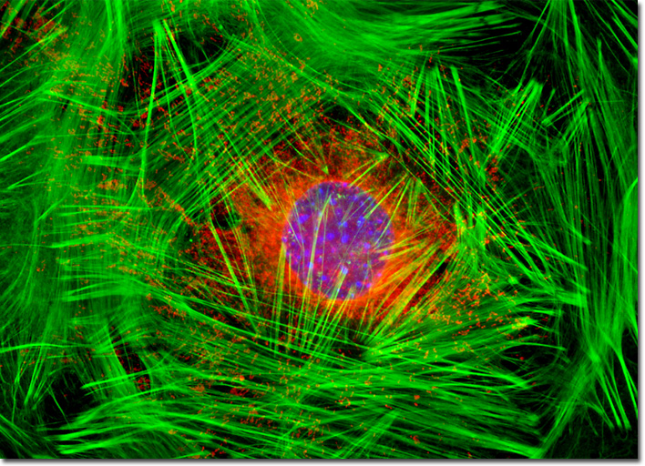
|
Similar to other connective tissue cells, fibroblasts develop in embryos from the mesenchyme, a network of loosely packed, unspecialized cells that have the potential to differentiate in several different ways depending upon various circumstances. Subsequent to embryonic development, it is generally believed that a small amount of mesenchymal cells survive in small blood vessels and other locations in the body, which can be called upon to differentiate into specialized cells when needed. In cases of injury, some of these mesenchymal cells may develop into fibroblasts in order to facilitate the healing of damaged tissues. In addition, fibroblasts that are already present in the area of the injury begin to grow in order to form a loose connective tissue structure over the damaged area. The prolific fibroblasts then produce substantial amounts of collagen, which helps provide strength to the developing tissue, eventually resulting in a scar that is almost solely comprised of the fibrous protein. The nuclei of embryonic Swiss mouse fibroblasts in culture (illustrated above) were targeted with the nucleic acid probe DAPI, which has an excitation maximum at 358 nanometers and an emission maximum at 461 nanometers when bound to DNA in cell cultures and tissue sections. In addition, the cells were also stained with Alexa Fluor 488 conjugated to phalloidin (filamentous actin) and MitoTracker Red CMXRos (mitochondria). Images were recorded in grayscale with a QImaging Retiga Fast-EXi camera system coupled to an Olympus BX-51 microscope equipped with bandpass emission fluorescence filter optical blocks provided by Omega Optical. During the processing stage, individual image channels were pseudocolored with RGB values corresponding to each of the fluorophore emission spectral profiles. |
© 1995-2025 by Michael W. Davidson and The Florida State University. All Rights Reserved. No images, graphics, software, scripts, or applets may be reproduced or used in any manner without permission from the copyright holders. Use of this website means you agree to all of the Legal Terms and Conditions set forth by the owners.
This website is maintained by our
|