Observing Mitosis with Fluorescence Microscopy
Metaphase
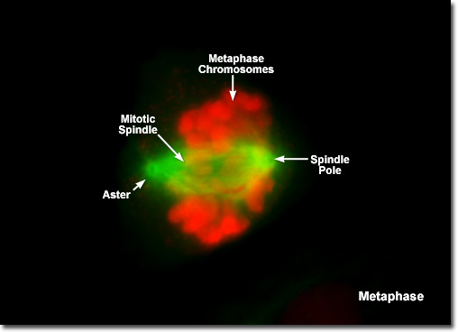
|
As the dividing cell approaches metaphase, the chromosomes become highly condensed and the mitotic spindle appears (visible as a series of green tubules in the digital images of PtK2 metaphase). In fluorescence images of mitosis, the two poles of the spindle and the spindle fibers are clearly visible, as are the asters, which radially migrate away from the metaphase plate. The mitotic spindle contains the fibers (microtubules) responsible for translocating and separating the chromosomes. The micrograph presented above captures a PtK2 rat kangaroo epithelial kidney cell in the transition from prometaphase to metaphase. The chromosomes were treated with primary antibodies to CDC6, a nuclear protein, and immunolabeled with secondary antibody fragments conjugated to Alexa Fluor 586 (red fluorescence). |
© 1995-2025 by Michael W. Davidson and The Florida State University. All Rights Reserved. No images, graphics, software, scripts, or applets may be reproduced or used in any manner without permission from the copyright holders. Use of this website means you agree to all of the Legal Terms and Conditions set forth by the owners.
This website is maintained by our
|