Observing Mitosis with Fluorescence Microscopy
Cytokinesis
|
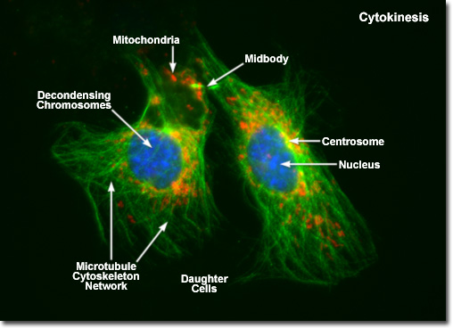
Presented in the digital fluorescence micrograph above is a pair of newly formed daughter rat kangaroo (PtK2) kidney epithelial cells in the late stages of cytokinesis. The chromatin is stained with a blue fluorescent probe (DAPI), while the cytoskeletal microtubule network (mitotic spindle) is stained green (Alexa Fluor 488) and cellular mitochondria are stained with a red dye (MitoTracker Red CMXRos). Note that the mitochondria are becoming interspersed throughout the cytoplasm, the previously condensed chromosomes are forming interphase chromatin, and the mitotic spindle is being redistributed into a cytoskeletal network. A thin bridge between the daughter cells, termed the midbody, is visible under the microscope for several hours before it finally disappears, leaving two completely independent daughter cells. |
© 1995-2022 by Michael W. Davidson and The Florida State University. All Rights Reserved. No images, graphics, software, scripts, or applets may be reproduced or used in any manner without permission from the copyright holders. Use of this website means you agree to all of the Legal Terms and Conditions set forth by the owners.
This website is maintained by our
|