Observing Mitosis with Fluorescence Microscopy
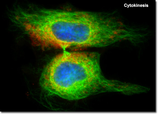
Cytokinesis
|
Orientation of the contractile ring and cleavage furrow, which lie parallel to the metaphase plate and perpendicular to the polar axis of the spindle, is strictly controlled by the mitotic apparatus during cytokinesis. In the microscope, the first visible sign of cleavage in animal cells is an inward folding, or furrowing, of the plasma membrane during late anaphase. A precise orientational relationship between the mitotic spindle and the cleavage furrow ensures that chromatids separated during anaphase will be evenly distributed into the two daughter cells during cell division, guaranteeing that both cells receive identical copies of the genetic material. |
© 1995-2025 by Michael W. Davidson and The Florida State University. All Rights Reserved. No images, graphics, software, scripts, or applets may be reproduced or used in any manner without permission from the copyright holders. Use of this website means you agree to all of the Legal Terms and Conditions set forth by the owners.
This website is maintained by our
|