Polarized Light Microscopy Digital Image Gallery
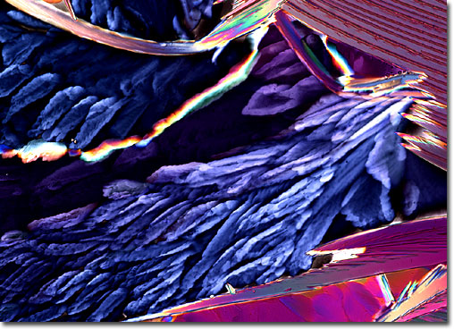
Safranin O
|
Safranin O is a stain frequently utilized in microscopy that appears as dark red to brown crystals at room temperature. According to most sources, safranin O is a mixture of the compounds dimethyl safranin and trimethyl safranin, though some only list the dimethyl compound as a constituent of the stain. Safranin O is most commonly employed for counterstaining nuclei red, but may also be used to stain chromosomes and cell walls. Similar to most substances found in the laboratory, safranin O must be handled with care. The stain can cause both skin and eye irritation, and may be harmful if swallowed, absorbed through the skin, or inhaled. Safranin O is also considered an irritant of mucous membranes and the upper respiratory tract, but is not flammable or explosive under typical laboratory conditions. |
© 1995-2022 by Michael W. Davidson and The Florida State University. All Rights Reserved. No images, graphics, software, scripts, or applets may be reproduced or used in any manner without permission from the copyright holders. Use of this website means you agree to all of the Legal Terms and Conditions set forth by the owners.
This website is maintained by our
|