
Polarized Light Microscopy Digital Image Gallery
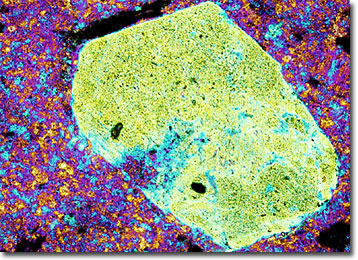
Rhyolite Porphyry
Some of the best-known examples of rhyolite can be found in the Yellowstone Park region and in the southwestern United States. Many of the rocks in those areas formed approximately 600,000 years ago through a series of massive volcanic eruptions.
Rhyolite is a fine-grained extrusive igneous rock that is mineralogically similar to granite. It chiefly contains orthoclase feldspar and quartz, but may contain lesser amounts of plagioclase feldspar, pyroxenes, biotite, and amphiboles. Some varieties of rhyolite are also similar to granite in appearance and may be easily mistaken for that rock in hand specimens. However, other types of rhyolite, such as the glassy obsidian, perlite, and pitchstone, are much more distinctive.
Relatively abundant around the world, rhyolites have been formed in all geologic ages. Most of the rocks are porphyritic in texture, consisting of a fine-grained base through which sizable crystals, called phenocrysts, are dispersed. The two different sizes of crystals in these rocks is an indication that the rhyolite cooled in two separate stages. Crystallization that begins when the magma is still buried deep within the Earth produces the phenocrysts, while the groundmass they are lodged in does not form until after an eruption brings the magma to the Earth’s surface.